 ISSN: 2822-0838 Online

The Validity of Iscan’s Age Estimation Method applied to the Fourth Rib in a Thai Male population
Phimolsinee Siriphimolwat, Watha Minsan, and Karnda Mekjaidee*Published Date : 2022-01-21
DOI : https://doi.org/10.12982/CMUJNS.2022.018
Journal Issues : Number 1, January-March 2022
Abstract Iscan’s staging method for age estimation from fourth ribs has been proved to be a potentially useful tool for various populations. However, due to interracial variations, it is necessary to calibrate its’ accuracy in reference to a population of Thai males before it can be applied. This study aimed to evaluate the validity of phasing analyses of sternal ends of fourth ribs developed by Iscan, et al., on a Thai male population. The Iscan’s method was applied to 50 Thai males aged 18 years and above. Staging of sternal ends of the ribs was analyzed based on 3 morphological features: pit depth, pit shape, and rim and wall configurations – designated as components 1, 2, and 3, respectively. The data was analyzed using descriptive statistics, one-way ANOVA, and cross-tabulation with Kendall’s Tau-c correlation. It was found that sternal end stages of fourth ribs in each component, inclusive of composite scores, are positively correlated to the age of the individual (P < 0.001). However, Iscan’s result - which was derived from a population of white males - delivers age-underestimation in a Thai population. Hence, we developed a modified age prediction reference table for application to Thai males. Applying cross-tabulation analysis, stages 1 and 2 were found only in individuals who were less than 40 years old, and stages 4 and 5 were found only in the 40 years and above age-group except for 1 case.
Keywords: Age estimation, Fourth rib, Iscan’s method, Thai male
Funding: This research did not receive grant from any funding agencies.
Citation: Siriphimolwat, P., Minsan, W., Mekjaidee, K. 2021. The validity of iscan’s age estimation method applied to the fourth rib in a Thai male population. CMU J. Nat. Sci. 21(1): e2022018.
INTRODUCTION
When a skeletonized body is found, it is necessary to gain primary information about its’ ancestry, sex, stature, and age, in order to review it against a list of possible missing persons. Pelvises and skulls are generally used for estimation of age at death. Accuracy varies, and is contingent upon forensic methods, as well as the age and ancestry of the individual. Key, et al., in 1994, reported that endocranial suture closures could be used for age estimation. Their results showed that the method worked well only in young and middle-age groups less than 50 years of age (Key, Aiello, and Molleson 1994). In 2004, Schmitt revealed a method of age estimation utilizing pubic symphyses and the auricular surfaces of ilia, which they applied to Thai samples. However, their efforts culminated in overestimation of the predicted ages (Schmitt 2004). This confirmed the hypothesis that inter-population differences exist, and before the adoption of any one method, pre-testing and calibrating for these differences in each population is needed.
Especially during the last decade, many methods of age prediction using bones and teeth have been investigated using Thai male populations. Most of the methods are based on changes in bone morphology through aging. The notorious skull suture closure method was studied by Ruengdit et al., in 2018. Thai skulls of both sexes presented a wide range of inaccurate data (Ruengdit et al. 2018).
Though they are not mentioned as frequently in the science of age prediction, vertebrae also have been reported as a useful tool for Thai populations. In 2018, Suwanlikhid et al., studied the degree of osteophyte formation in 125 male lumbar spines, and found it was capable of providing age estimation (Suwanlikhid et al. 2018) Later, Praneatpolgrang et al., developed an age-estimation equation incorporating osteophyte length in adult Thai males (Praneatpolgrang, Prasitwattanaseree, and Mahakkanukrauh 2019). Femur bone has also been reported as useful. In 2017, Khomkham et al., found 3 features of the acetabulum that might be useful in the age estimation of Thai males (Khomkham et al. 2017).
The histomorphology of bones has been explored and found to be helpful in age estimation. In 2019, Chompoophuenl et al., developed a predictive model of age estimation for Thai males using 3 histological variables: the perimeter of the Haversian canal, the percentage of lamellar bone area, and the collagen measured using an image processing technique and the MATLAB program. The correlation coefficient of their equation was high (Chompoophuenl et al. Jun2019). Further histological study of Thai male lumbar spines was also done regarding age estimation. Sattarath et al., in 2021, reported a negative relationship between the percentage of the trabecular area over the total area and age of male individuals (Sattarath et al. 2021).
Imaging of bones has also proven to be useful for the age prediction of Thai males. To this end, Pattamapaspong et al., in 2014, performed staging analysis of the ossification of medial clavicular epiphyses using thin-sliced computed tomography on both male and female Thai patients (Pattamapaspong et al. 2015). Chest plate radiographic images of 136 Thai males were inspected by Monum et al., in 2017. They proved that the presence of any ossification indicated an age greater than 29 years old and established a model for calculate the age of the individual (Monum et al. 2017).
In addition to bones, teeth have been reported to have a good correlation with age in Thai populations. Duangto et al., in 2017, investigated third molars, and found high accuracy in their ability to predict age for both males and females (Duangto et al. 2017).
Therefore, the existence of such an abundance of methods that can predict age at death in Thai males makes it more likely that greater accuracy in these predictions can be achieved. Since not every remains found at the scene of an investigation is complete body, a greater number of more varied approaches increases the odds of identifying the individuals involved.
Rib bones have been reported to benefit age estimation in males. In 1984, Iscan derived a new method of morphological phasing analysis for age determination using the right fourth ribs from 93 white males. Three feature components of the sternal ends of these ribs were examined: pit depth (component 1), pit shape (component 2), and rim and wall configurations (component 3). Each component was divided into 6 stages. The results indicated that this method was very effective at determining age (İşcan, Loth, and Wright 1984). Iscan’s method was thereafter applied to varied populations and has been found to be very effective (Işcan, Loth, and Wright 1987; Yavuz, İşcan, and Çöloğlu 1998; Oettlé and Steyn 2000; Meena et al. 2012; Zahra et al., n.d.; Haj Salem et al. 2014; Cerezo-Román and Hernández Espinoza 2014; Muñoz et al. 2018). As far as the authors are aware, there have been no morphological studies of fourth ribs using Iscan’s method to determine age in a Thai population. Pretesting before applying to Thai males is necessary because of the interracial variation.
The current study’s aim was to evaluate the validity of Iscan’s age estimation method using morphological phase analysis of fourth ribs’ sternal ends in Thai male population.
MATERIAL AND METHODS
The samples came from a group of 50 Thai males whose ages ranged from 18 to 89 years. All were autopsied at the department of forensic medicine, faculty of medicine, at Chiang Mai University. All samples were collected after obtaining permission from the families of the deceased. This study was approved as adhering to the ethical standards as dictated by the research ethics committee (Research ID: 4727/Study code: NONE-2560-04727) of the Faculty of Medicine, Chiang Mai University. Age distribution of the samples is shown in Table 1.
Table 1. Age distribution of the samples.
|
Age intervals (yrs.) |
N |
% |
|
0-16 |
0 |
0.00 |
|
17-19 |
4 |
8.00 |
|
20-29 |
8 |
16.00 |
|
30-39 |
6 |
12.00 |
|
40-49 |
6 |
12.00 |
|
50-59 |
10 |
20.00 |
|
60-69 |
6 |
12.00 |
|
70 and over |
10 |
20.00 |
|
Total |
50 |
100.00 |
Sternal ends of the right fourth ribs were removed and soaked in water for 3 days. On day 4 they were boiled in water mixed with simple laundry detergent at 60 degrees for 4 hours. The ratio of detergent to water was 30gm: 3L. Next, soft tissue was scraped from the ribs by using wooden tongue depressors. The ribs were continuously soaked in water, and the process repeated, until all soft tissue was successfully removed. All ribs were completely cleaned after soaking and boiling for 4-7 days. After the soft tissue was removed, the ribs were left to dry at room temperature for one day, then staged according to Iscan’s method.
Reviewing the 3 morphological features (components 1, 2, and 3), the sternal ends of the ribs were subjected to stage analysis per Iscan’s method. Component 1 was
pit depth; component 2 was pit shape; component 3 was rim and wall configurations (Figure 1).
Component 1 is divided into 6 stages as following:
Stage 0 Flat or slightly undulated with depth not more than 1.1 mm.
Stage 1 Well-seen pit with depth ranges from 1.1 and 2.5 mm.
Stage 2 Pit depth from 2.6 to 4.5 mm.
Stage 3 Pit depth from 4.6 to 7.0 mm.
Stage 4 Pit depth from 7.1 to 10.0 mm.
Stage 5 Pit depth greater than 10.0 mm.
Component 2 is divided into 6 stages as following:
Stage 0 No pit. Flat or slightly undulated at medial articular surface.
Stage 1 Well-seen pit with clear anterior and posterior wall.
Stage 2 V-shaped with a thick wall.
Stage 3 Narrow U-shaped with a thick wall.
Stage 4 Wide U-shaped with a thin wall.
Stage 5 Wide U-shaped. Wall is deeper, more brittle, and poorer in texture.
Component 3 is divided into 6 stages as following:
Stage 0 Smooth edge with no wall.
Stage 1 Smooth edge with a thick wall.
Stage 2 Smooth and scalloped edge with a thick wall.
Stage 3 Rough edge with a thin and smooth wall.
Stage 4 Sharp and jagged edge. Wall is thinner and has a deteriorated texture.
Stage 5 Sharp and brittle edge with long bony projections. Wall is incomplete.
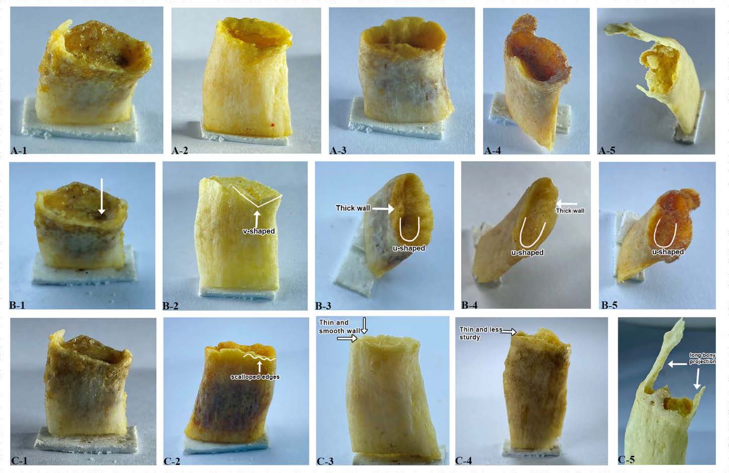
Figure 1. Stages of sternal ends of fourth ribs according to Iscan’s method. A, B and C represent components 1, 2, and 3. Numbers 1 to 5 represent stages 1 through 5 of each component.
Composite staging scores were calculated by adding stages or scores to each component. The data was analyzed using descriptive statistics, one-way ANOVA, and cross-tabulation with Kendall’s Tau-c correlation. All statistical analysis was done by using SPSS 20.
RESULTS
Descriptive analysis of the chronological ages of the 50 samples was performed. The youngest and the oldest samples were 18 and 89 years old, respectively. The mean age of the samples was 49.7 years, and the standard deviation was 21.484 years. The data set had a normal distribution, and exhibited perfect skewness, and an acceptable kurtosis.
Table 2 contains descriptive statistics of the mean ages of Thai males compared to white males from Iscan’s study. For Thai males, the mean age in all components increased less than 10 years from stage 1 to stage 2; then, increases of 15-19, 10-23 and 11-23 years were found from stages 2 through 5 for components 1, 2 and 3, respectively. The 95% confidence interval of every stage in each component is narrow, suggesting that prominent changes occurred throughout the subjects’ lifetimes. This finding contrasts with Iscan’s study, in which it was showed that the change occurred rapidly only at the age of 30 years or less. Later ages showed the change was not as generous (İşcan, Loth, and Wright 1984). Scatter plots of the samples appear in Figure 2. The correlations are positive lineage specific.
Table 2. Descriptive statistics of the mean ages of Thai males and white males in Iscan’s study.
|
Stages or scores |
Thai males (This study) |
|
White males (Iscan’s study) |
||||||||
|
N |
Mean age |
SD |
95% Confidence interval of mean |
Age range |
|
N |
Mean age |
SD |
95% Confidence interval of mean |
Age range |
|
|
Component 1 |
|
|
|
|
|
|
|
|
|
|
|
|
1 |
4 |
19.50 |
1.29 |
17.45 – 21.55 |
18 – 21 |
|
9 |
20.30 |
3.32 |
17.80 - 22.90 |
17 – 25 |
|
2 |
13 |
28.38 |
6.28 |
24.59 – 32.18 |
19 – 37 |
|
29 |
30.70 |
12.40 |
26.00 - 35.40 |
18 – 64 |
|
3 |
10 |
47.50 |
9.41 |
40.77 – 54.23 |
27 – 58 |
|
31 |
40.90 |
13.72 |
35.80 - 46.00 |
21 – 67 |
|
4 |
19 |
65.21 |
13.28 |
58.81 – 71.61 |
45 – 89 |
|
9 |
55.00 |
15.39 |
43.20 - 66.80 |
32 – 76 |
|
5 |
4 |
81.00 |
5.03 |
72.99 – 89.01 |
76 – 89 |
|
4 |
57.50 |
12.92 |
36.90 - 78.10 |
44 – 70 |
|
Total |
50 |
48.32 |
7.06 |
42.92 – 53.72 |
18 - 89 |
|
82 |
37.90 |
16.15 |
34.80 – 40.90 |
17 – 85 |
|
Component 2 |
|
|
|
|
|
|
|
|
|
|
|
|
1 |
5 |
19.80 |
1.64 |
17.76 – 21.84 |
18 – 22 |
|
4 |
17.30 |
0.50 |
16.50 - 18.00 |
17 – 18 |
|
2 |
5 |
23.20 |
3.70 |
18.60 – 27.80 |
19 – 28 |
|
15 |
22.80 |
3.28 |
21.00 - 24.60 |
18 – 30 |
|
3 |
7 |
33.14 |
3.34 |
30.06 – 36.23 |
28 – 37 |
|
28 |
30.50 |
9.61 |
26.80 - 34.30 |
19 – 66 |
|
4 |
17 |
50.41 |
9.28 |
45.64 – 55.18 |
27 – 66 |
|
22 |
47.10 |
11.61 |
41.90 - 52.20 |
26 – 67 |
|
5 |
16 |
73.81 |
11.24 |
67.82 – 79.80 |
55 – 89 |
|
15 |
61.60 |
12.94 |
54.40 - 68.80 |
44 – 85 |
|
Total |
50 |
40.07 |
5.84 |
35.98 – 44.17 |
18 – 89 |
|
84 |
38.40 |
17.26 |
34.70 – 42.20 |
17 – 85 |
|
Component 3 |
|
|
|
|
|
|
|
|
|
|
|
|
1 |
6 |
19.33 |
1.03 |
18.25 – 20.42 |
18 – 21 |
|
5 |
17.80 |
1.30 |
16.20 - 19.40 |
17 – 20 |
|
2 |
6 |
26.67 |
3.20 |
23.30 – 30.03 |
22 – 31 |
|
25 |
24.10 |
3.55 |
22.70 - 25.60 |
18 – 31 |
|
3 |
8 |
38.13 |
10.32 |
29.50 – 46.75 |
25 – 56 |
|
20 |
34.30 |
11.62 |
28.90 - 39.70 |
21 – 66 |
|
4 |
17 |
53.41 |
8.38 |
49.10 – 57.72 |
42 – 68 |
|
16 |
49.50 |
11.21 |
43.50 - 55.50 |
32 – 71 |
|
5 |
13 |
76.62 |
10.21 |
70.45 – 82.79 |
60 - 89 |
|
16 |
58.20 |
11.53 |
52.00 - 64.30 |
43 – 76 |
|
Total |
50 |
42.83 |
6.63 |
38.12 – 47.54 |
18 - 89 |
|
82 |
37.80 |
16.67 |
34.20 – 41.50 |
17 – 76 |
|
Composite scores |
|
|
|
|
|
|
|
|
|
||
|
3 |
3 |
19.33 |
1.53 |
15.54 – 23.13 |
18 - 21 |
|
3 |
17.00 |
0.00 |
17.00 – 17.00 |
17 - 17 |
|
4 |
2 |
19.50 |
0.71 |
13.15 – 25.85 |
19 – 20 |
|
2 |
19.00 |
1.41 |
17.00 – 31.70 |
18 - 20 |
|
5 |
2 |
20.50 |
2.12 |
1.44 – 39.56 |
19 – 22 |
|
4 |
22.50 |
3.32 |
17.20 – 27.80 |
18 – 25 |
|
6 |
2 |
26.00 |
2.83 |
0.59 – 51.41 |
24 – 28 |
|
7 |
23.10 |
4.06 |
19.40 – 26.90 |
18 – 30 |
|
7 |
3 |
28.00 |
3.00 |
20.55 – 35.45 |
25 – 31 |
|
12 |
24.90 |
3.63 |
22.60 - 27.20 |
19 – 31 |
|
8 |
5 |
34.60 |
2.51 |
31.48 – 37.72 |
31 – 37 |
|
9 |
27.00 |
4.90 |
23.20 – 30.80 |
21 – 36 |
|
9 |
1 |
27.00 |
- |
- |
27 |
|
10 |
37.80 |
13.21 |
28.30 - 47.30 |
24 – 66 |
|
10 |
2 |
53.50 |
3.54 |
21.73 – 85.27 |
51 – 56 |
|
8 |
47.10 |
12.03 |
37.10 – 57.20 |
30 – 64 |
|
11 |
6 |
47.17 |
6.01 |
40.86 – 53.48 |
42 – 58 |
|
6 |
48.50 |
9.89 |
38.10 – 58.80 |
41 – 67 |
|
12 |
8 |
54.75 |
7.25 |
48.69 – 60.81 |
45 – 66 |
|
7 |
47.60 |
11.75 |
36.70 – 58.40 |
32 – 67 |
|
13 |
4 |
61.75 |
5.56 |
52.90 – 70.60 |
55 – 68 |
|
5 |
56.00 |
10.32 |
43.20 – 68.80 |
44 – 71 |
|
14 |
8 |
76.50 |
10.84 |
67.44 – 85.56 |
61 – 89 |
|
4 |
63.50 |
12.26 |
44.00 – 83.00 |
52 – 76 |
|
15 |
4 |
81.00 |
5.03 |
72.99 – 89.01 |
76 – 88 |
|
4 |
57.50 |
12.92 |
36.90 – 78.10 |
44 – 70 |
|
Total |
50 |
40.20 |
3.92 |
29.80 – 50.60 |
18 - 89 |
|
84 |
37.30 |
16.81 |
33.80 – 41.00 |
17 – 76 |
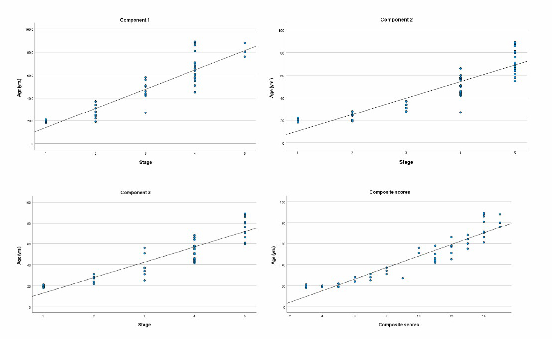
Figure 2. Scatter plots of the 3 components and composite scores.
Statistical analysis of variance (one-way ANOVA), along with effect size, in terms of eta-squared (η2), is shown in Table 3. The F-ratio indicates significant association between chronological age and stage in every component, including composite scores at the level of P < 0.001. η2 reveals that 80%, 85%, 86% and 93% of variation in age can be explained by the stages of components 1, 2, and 3, and composite scores, which are higher than those in Iscan’s study (41%, 68%, 70%, and 73% respectively).
Table 3. One-way analysis of variance of the 3 components and composite scores.
|
Sources of variation |
Sum of squares |
d.f. |
Mean squares |
F-ratio |
η2 |
|
|
Component 1 |
|
|||||
|
Between groups |
18,092.77 |
4 |
4,523.19 |
44.99* |
0.80 |
|
|
Within groups |
4,523.74 |
45 |
100.53 |
|
|
|
|
Total |
22,616.50 |
49 |
|
|
|
|
|
Component 2 |
|
|||||
|
Between groups |
19,211.49 |
4 |
4,802.87 |
63.47* |
0.85 |
|
|
Within groups |
3,405.01 |
45 |
75.67 |
|
|
|
|
Total |
22,616.50 |
49 |
|
|
|
|
|
Component 3 |
|
|||||
|
Between groups |
19,439.76 |
4 |
4,859.94 |
68.84* |
0.86 |
|
|
Within groups |
3,176.74 |
45 |
70.59 |
|
|
|
|
Total |
22,616.50 |
49 |
|
|
|
|
|
Composite scores |
|
|||||
|
Between groups |
21,004.05 |
12 |
1,750.34 |
40.16* |
0.93 |
|
|
Within groups |
1,612.45 |
37 |
43.58 |
|
|
|
|
Total |
22,616.50 |
49 |
|
|
|
|
Note: *Significant at P < 0.001 level
Testing for multiple comparisons of mean age was done by using the Least Significant Difference (LSD). Significant differences existed between every stage, and among all components, except for between stage 1 and 2 in every component, and stage 2 and 3 in component 2 (Table 4). A post hoc test was not performed for composite scores because there were fewer than 2 cases in at least one group.
Table 4. LSD test for multiple comparisons of mean age.
|
Stages |
Compared |
|
Mean Difference |
|
|
Component 1 |
Component 2 |
Component 3 |
||
|
1 |
2 |
-8.885 |
-3.400 |
-7.333 |
|
|
3 |
-28.000* |
-13.343* |
-18.792* |
|
|
4 |
-45.711* |
-30.612* |
-34.078* |
|
|
5 |
-61.500* |
-54.012* |
-57.282* |
|
2 |
1 |
8.885 |
3.400 |
7.333 |
|
|
3 |
-19.115* |
-9.943 |
-11.458* |
|
|
4 |
-36.826* |
-27.212* |
-26.745* |
|
|
5 |
-52.615* |
-50.612* |
-49.949* |
|
3 |
1 |
28.000* |
13.343* |
18.792* |
|
|
2 |
19.115* |
9.943 |
11.458* |
|
|
4 |
-17.711* |
-17.269* |
-15.287* |
|
|
5 |
-33.500* |
-40.670* |
-38.490* |
|
4 |
1 |
45.711* |
30.612* |
34.078* |
|
|
2 |
36.826* |
27.212* |
26.745* |
|
|
3 |
17.711* |
17.269* |
15.287* |
|
|
5 |
-15.789* |
-23.401* |
-23.204* |
|
5 |
1 |
61.500* |
54.013* |
57.282* |
|
|
2 |
52.615* |
50.613* |
49.949* |
|
|
3 |
33.500* |
40.670* |
38.490* |
|
|
4 |
15.789* |
23.401* |
23.204* |
Note: *The mean difference is significant at < 0.001
Cross-tabulation for frequency distribution according to age intervals and stages of each component was analyzed (Table 5). It was noted that stages 1 and 2 in all three components were not found in samples greater than 39 years old, and stages 4 and 5 were not found in samples less than 40 years old, except for 1 case in which component 2 was found in the 20-29 years old group. The composite scores could be divided into 2 groups: scores 3-9 vs. scores 10-15. The former group included members aged less than 40 years, and the latter had members 40 years old and above.
Table 5. Frequency distribution of stages in 3 components by age intervals
|
Stages |
Age intervals (years) |
|
|||||||||||||||||
|
17-19 |
20-29 |
30-39 |
40-49 |
50-59 |
60-69 |
370 |
Total N |
||||||||||||
|
Component 1a |
|
|
|
|
|
|
|
|
|||||||||||
|
1 |
2 |
2 |
|
|
|
|
|
4 |
|||||||||||
|
2 |
2 |
5 |
6 |
|
|
|
|
13 |
|||||||||||
|
3 |
|
1 |
|
4 |
5 |
|
|
10 |
|||||||||||
|
4 |
|
|
|
2 |
5 |
6 |
6 |
19 |
|||||||||||
|
5 |
|
|
|
|
|
|
4 |
4 |
|||||||||||
|
Total N |
4 |
8 |
6 |
6 |
10 |
6 |
10 |
50 |
|||||||||||
|
Component 2b |
|
|
|
|
|
|
|
|
|||||||||||
|
1 |
3 |
2 |
|
|
|
|
|
5 |
|||||||||||
|
2 |
1 |
4 |
|
|
|
|
|
5 |
|||||||||||
|
3 |
|
1 |
6 |
|
|
|
|
7 |
|||||||||||
|
4 |
|
1 |
|
6 |
8 |
2 |
|
17 |
|||||||||||
|
5 |
|
|
|
|
2 |
4 |
10 |
16 |
|||||||||||
|
Total N |
4 |
8 |
6 |
6 |
10 |
6 |
10 |
50 |
|||||||||||
|
Stages |
Age intervals (years) Age intervals (years) Age intervals (years) Age intervals (years) |
|
|||||||||||||||||
|
17-19 |
20-29 |
30-39 |
40-49 |
50-59 |
60-69 |
370 |
Total N |
|
|||||||||||
|
Component 3c |
|
|
|
|
|
|
|
|
|||||||||||
|
1 |
4 |
2 |
|
|
|
|
|
6 |
|||||||||||
|
2 |
|
5 |
1 |
|
|
|
|
6 |
|||||||||||
|
3 |
|
1 |
5 |
|
2 |
|
|
8 |
|||||||||||
|
4 |
|
|
|
6 |
8 |
3 |
|
17 |
|||||||||||
|
5 |
|
|
|
|
|
3 |
10 |
13 |
|||||||||||
|
Total N |
4 |
8 |
6 |
6 |
10 |
6 |
10 |
50 |
|||||||||||
|
Composite scoresd |
|
|
|
|
|
|
|
|
|||||||||||
|
3 |
2 |
1 |
|
|
|
|
|
3 |
|||||||||||
|
4 |
1 |
1 |
|
|
|
|
|
2 |
|||||||||||
|
5 |
1 |
1 |
|
|
|
|
|
2 |
|||||||||||
|
6 |
|
2 |
|
|
|
|
|
2 |
|||||||||||
|
7 |
|
2 |
1 |
|
|
|
|
3 |
|||||||||||
|
8 |
|
|
5 |
|
|
|
|
5 |
|||||||||||
|
9 |
|
1 |
|
|
|
|
|
1 |
|||||||||||
|
10 |
|
|
|
|
2 |
|
|
2 |
|||||||||||
|
11 |
|
|
|
4 |
2 |
|
|
6 |
|||||||||||
|
12 |
|
|
|
2 |
5 |
1 |
|
8 |
|||||||||||
|
13 |
|
|
|
|
1 |
3 |
|
4 |
|||||||||||
|
14 |
|
|
|
|
|
2 |
6 |
8 |
|||||||||||
|
15 |
|
|
|
|
|
|
4 |
4 |
|||||||||||
|
Total N |
4 |
8 |
6 |
6 |
10 |
6 |
10 |
50 |
|||||||||||
Note: aKendall’s Tau-c correlations = 0.801 (P < 0.001), bKendall’s Tau-c correlations = 0.856 (P < 0.001), cKendall’s Tau-c correlations = 0.880 (P < 0.001), dKendall’s Tau-c correlations = 0.894 (P < 0.001)
The kappa statistic was used to test inter-rater reliability. Ten samples were staged by the researcher, and a master’s student in forensic science, and a PhD student in forensic osteology. The interrater agreement measurements for components 1, 2, and 3, were 1, 0.8, and 0.8, respectively.
DISCUSSION
Phasing analysis of the sternal ends of fourth ribs developed by Iscan, et al., is an effective method for estimating age at death. Nevertheless, considering the white ancestry of the individuals whose samples were recruited for Iscan’s study, a wider ranging validity of the method needs to be proved before applying it to non-white populations.
Testing in Turkish, Indian, and Pakistani populations gave the same results as in the white populations (Yavuz, İşcan, and Çöloğlu 1998; Meena et al. 2012; Zahra et al. 2020). However, there are some reports of less accuracy when applied to some other kinds of populations. In 1987, Iscan tested his own method in 53 African American males and found that there were morphological and chronological differences in the samples with ages greater than early 30 years (Işcan, Loth, and Wright 1987). In 2000, Oettlé et al., applied the method to 265 South African black male skeletons. They found that the repeatability of the method was acceptable, but that accuracy in age prediction was reduced (Oettlé and Steyn 2000). Salem et al., applied the method to a Tunisian population in 2014, and found that the assessed and actual ages were only well-related in people aged less than 39 years old (Haj Salem et al. 2014). Cerezo-Roman and Espinoza applied it to Mexican males in 2014, and it yielded ages that were underestimated, especially when the age at death was greater than 40 years old (Cerezo-Román and Hernández Espinoza 2014). In 2018, Muñoz et al., tested the validity of Iscan’s method on 444 male skeletons in Mexico City and found good correlation, yet it underestimated age at death, and inaccuracy increased at higher stages (Muñoz et al. 2018).
This present study was done on a Thai population from an Asian country. The results show that Iscan’s method for age prediction is valid when applied to Thai males. Nevertheless, inquiry into the differences between populations revealed that the morphological changes at the sternal end of the fourth rib is, as aging occurs, more accelerated in Thais than in whites. Therefore, age prediction using Iscan’s own reference table yields an underestimated age of death, which confirms the existence of inter-population inaccuracy. For more accurate age estimation, this study provides a reference table for Thai males (Table 2). According to negative statistical differences between stages 1 and 2 of every component (Table 4), it is possible to define the age at death as less than 40 years old, if the fourth rib morphological change is in stage 1 or 2. The cross-tabulation of the distribution of stages by age intervals (Table 5) reveals an increase of stages with age. Simple formulas could be practically applied to cases on scene as follows: (1) stages 1 or 2 of any component reveal the age of the individual as less than 40 years old; (2) stages 4 or 5 signal age at 40 years old or above; and, (3) a composite score of 10 is the dividing line between ages less than and more than 40 years old.
Because of the high correlation value of every component (0.801, 0.856, and 0.880 for components 1, 2, and 3), any component could be confidently used in and of itself for age estimation. This is beneficial for cases in which there are incomplete sternal rib ends. The measures of interrater agreement by Kappa statistics were high (1, 0.8, and 0.8 for components 1, 2, and 3), indicating repeatability of the method.
The bone preparation period in this study was abbreviated compared to several studies including Iscan’s original method (İşcan, Loth, and Wright 1984; Yavuz, İşcan, and Çöloğlu 1998; Oettlé and Steyn 2000; Haj Salem et al. 2014). The bone cleaning technique for those studies was to let the bones decompose naturally, or let them soak in water, for several weeks; then finally to boil them in water. It was reported that detergent was an effective, safe, and fast method for bone cleaning. The action of detergent at 40-60°C produced satisfactory result for forensic practice (Mairs, Swift, and Rutty 2004). After using detergent some have reported deterioration of tool marks (Uhre et al. 2015), and creation of porous bone surfaces (Lai et al. 2015). However, we did not include bone surface as criteria for ascertaining stages in this study.
The main restraint on the present study is the limited sample size. A study with more male samples, and inclusive of Thai females, is suggested in order to establish more accurate and more useful results from forensic application to Thai skeletons. Moreover, in regard to the greater application of radiography in current forensic anthropology, computed tomography (CT) scans could be used to get morphological images of the fourth rib instead of employing direct examination. CT scans can reduce working time because neither bone preparation nor dissection of the body is needed. The benefits of such an approach have been proven in Indian and Australian populations (Merritt 2018; Blaszkowska, Flavel, and Franklin 2019).
CONCLUSION
Age estimation in adults remains a developing but challenging task in forensic practice. As more methods are applied, age at death predictions become more accurate. Phase analysis of sternal ends of fourth ribs in a Thai male population was performed by using Iscan’s method. The 3 components indicating morphological change are pit depth, pit shape, and configurations of the rim and wall. It is viable to use Iscan’s method to ascertain age prediction in Thai males from stage analysis of the sternal end of the fourth rib. However, Iscan’s own reference yields underestimated age predictions in Thais. Therefore, a modification of the reference was developed based on the Thai male samples used in this study. This study delivers an age prediction reference for Thai males in table 3, and a practical application of it that can easily be used at investigatory scenes. Further explorations with larger sample sizes, with female-inclusive data groups, will be more constructive for divining age at death in Thai populations.
ACKNOWLEDGEMENTS
The authors would like to thank the relatives of the deceased for their permission to collect the samples. We would also like to thank the staff of the Department of Forensic Medicine, Faculty of Medicine, Chiang Mai University for their cooperation and support.
AUTHOR CONTRIBUTIONS
Phimolsinee Siriphimolwat designed, conducted the experiments, performed statistical analysis, and contributed to writing the manuscript. Watha Minsan contributed to statistical analysis and writing the manuscript. Karnda Mekjaidee contributed to statistical analysis, designed, and writing the manuscript. All authors have read and approved the final manuscript.
CONFLICT OF INTEREST
The authors declare no competing interest.
REFERENCES
Blaszkowska, M., Flavel, A., and Franklin, D. 2019. Validation of the işcan method in clinical MSCT scans specific to an australian population. International Journal of Legal Medicine 133: 1903–13.
Cerezo-Román, J.I., and Espinoza, P.O. 2014. Estimating age at death using the sternal end of the fourth ribs from mexican males. Forensic Science International. 236: 196.e1-196.e6.
Chompoophuenl, H., Mahakkanukrauh, P., Settakorn, J., Mekjaidee, K., Prasitwattanaseree, S., and Thumthong, W.J. 2019. Image processing technique for age estimation in thai adults by histomorphometry of decalcified cortical bone. International Medical Journal 26: 209–12.
Duangto, P., Iamaroon, A., Prasitwattanaseree, S., Mahakkanukrauh, P., and Janhom, A. 2017. New Models for Age Estimation and Assessment of Their Accuracy Using Developing Mandibular Third Molar Teeth in a Thai Population. International Journal of Legal Medicine 131: 559–68.
Işcan, M.Y., Loth, S.R., and Wright R.K. 1987. Racial Variation in the Sternal Extremity of the Rib and Its Effect on Age Determination. Journal of Forensic Sciences. 32: 452–66.
İşcan, M.Y., Susan R. Loth, S.R., and Wright, R.K. 1984. Metamorphosis at the Sternal Rib End: A New Method to Estimate Age at Death in White Males. American Journal of Physical Anthropology 65: 147–56.
Key, C.A., Aiello, L.C., and Molleson, T. 1994. Cranial Suture Closure and Its Implications for Age Estimation. International Journal of Osteoarchaeology 4: 193–207.
Meena, M.C., Rani, Y., Naik, S.K., and Murari, A. 2012. Age estimation from the iv rib by phase analysis in Indian males. Australian Journal of Forensic Sciences 44: 261–71.
Merritt, C. E. 2018. Part I – Adult skeletal age estimation using ct scans of cadavers: revision of the fourth rib methods. Journal of Forensic Radiology and Imaging 14 (September): 39–49.
Monum T, Mekjaidee K, Pattamapaspong N, Prasitwattanaseree S. Age estimation by chest plate radiographs in a Thai male population. Science and Justice, 2017; 57: 169-173.
Muñoz, A., Maestro, N., Benito, M., Sánchez, J.A., Márquez-Grant, N., Trejo, D., and Ríos, L. 2018. Sex and age at death estimation from the sternal end of the fourth rib. Does Íşcan’s Method Really Work? Legal Medicine. 31: 24–29.
Oettlé, A.C., and Steyn, M. 2000. Age Estimation from Sternal Ends of Ribs by Phase Analysis in South African Blacks. Journal of Forensic Sciences. Journal of Forensic Sciences. 45: 1071–79.
Khomkham, P., Chotecharnont,W., Srinuan, P., Suriyasathaporn, J., Srisaikaew, P., Inchai, C., Mann, R., and Mahakkanukrauh, P. 2017. Association between Age and Acetabulum Morphological Changes in Dry Bones in the Thai Population. Chiang Mai Medical Journal 56: 21–28.
Pattamapaspong, N., Madla, C., Mekjaidee, K., and Namwongprom, S. 2015. Age estimation of a Thai population based on maturation of the medial clavicular epiphysis using computed tomography. Forensic Science International 246: 123.e1-123.e5.
Sattarath, P., Wantanajittikul, K., Prasitwattanaseree, S., Settakorn, J., and Mekjaidee, K. 2021. Age related lumbar trabecular bone in a Thai population. Chiang Mai University Journal of Natural Sciences, 20.
Praneatpolgrang, S., Prasitwattanaseree, S., and Mahakkanukrauh, P. 2019. Age estimation equations using vertebral osteophyte formation in a thai population: comparison and modified osteophyte scoring method. Anatomy & Cell Biology 52: 149.
Ruengdit, S., Prasitwattanaseree, S., Mekjaidee, K., Sinthubua, A., and Mahakkanukrauh, P. 2018. Age estimation approaches using cranial suture closure: a validation study on a Thai population. Journal of Forensic and Legal Medicine 53: 79–86.
Salem, N.H., Aissaoui, A., Mesrati, M.A., Belhadj, M., Quatrehomme, G., and Chadly, A. 2014. Age Estimation from the Sternal End of the Fourth Rib: A Study of the Validity of İşcan’s Method in Tunisian Male Population. Legal Medicine. 16: 385–89.
Schmitt, A. 2004. Age-at-death assessment using the os pubis and the auricular surface of the ilium: a test on an identified Asian sample. International Journal of Osteoarchaeology, 14: 1–6.
Simon, M., Swift, B., and Rutty, G.N. 2004. Detergent: An Alternative Approach to Traditional Bone Cleaning Methods for Forensic Practice. American Journal of Forensic Medicine & Pathology, 25: 276–84.
Soon, L.P., Khoo, L.S., MOHD HILMI, S., AHMAD HAFIZAM, H., MOHD SHAH, M., NURLIZA, A., and NAZNI, W. A. 2015. Effectiveness of Bone Cleaning Process Using Chemical and Entomology Approaches: Time and Cost. Malaysian Journal of Pathology, 37: 123–35.
Suwanlikhid, N., Prasitwattanaseree, S., Palee, P., and Mahakkanukrauh, P. 2018. Age estimation of lumbar vertebrae by visual assessment in a Thai population. La Clinica Terapeutica 169: e204–12.
Uhre, M.L., Eriksen, A.K., Simonsen, K.P., Rasmussen, A.R., Hjort, B.B., and Lynnerup, N. 2015. Enzymatic maceration of bone: a gentler technique than boiling. Medicine, Science and the Law 55: 90–96.
Yavuz, M.F., İşcan, M.Y., and Çöloğlu, A.S. 1998. Age assessment by rib phase analysis in turks. Forensic Science International. 98: 47–54.
Zahra, K., Shahid, M., Kazmi, S., and Malik, A.R. 2020. Age estimation from macroscopically examining sternal end of right fourth rib by using pit depth in deceased males. Pakistan Journal of Medical & Health Sciences. 14: 1661-1665.
OPEN access freely available online
Chiang Mai University Journal of Natural Sciences [ISSN 16851994]
Chiang Mai University, Thailand
https://cmuj.cmu.ac.th
Phimolsinee Siriphimolwat1, Watha Minsan2, and Karnda Mekjaidee3,*
1 Forensic science, Chiangmai University, Chiang Mai, 50200 Thailand
2 Department of Statistics, Faculty of Science, Chiang Mai University, Chiang Mai, 50200 Thailand
3 Department of Forensic medicine, Faculty of Medicine, Chiang Mai University, Chiang Mai, 50200 Thailand
Corresponding author: Karnda Mekjaidee, E-mail: Karnda.me@gmail.com
Total Article Views
Editor: Korakot Nganvongpanit,
Chiang Mai University, Thailand
Article history:
Received: October 11 2021;
Revised: November 11, 2021;
Accepted: November 18, 2021