 ISSN: 2822-0838 Online

Age Related Lumbar Trabecular Bone in a Thai Population
Pornpath Sattarath, Kittichai Wantanajittikul, Sukon Prasitwattanaseree, Jongkolnee Settakorn, and Karnda Mekjaidee*Published Date : 2021-04-30
DOI : https://doi.org/10.12982/CMUJNS.2021.069
Journal Issues : Number 3, July-September 2021
Abstract Within spinal column, the lumbar vertebrae are the most durable and usually left behind in severely burnt body. European studies have reported that these vertebrae are useful for age estimation. However, it is widely acknowledged that different ancestry necessitates different methods and includes a range of variables, therefore a study specific to Thai population is needed for accuracy in the identification of Thai individuals. To investigate the correlation between lumbar vertebrae, and age of the individual, L1-L5 drilled out from 75 Thai cadavers. After undergoing histological processing each slide was photographed. The images were processed using an image processing technique to calculate the percentage of trabecular bone area over total area (%TBA/TA). Using the Statistical Package for the Social Sciences (SPSS) program, %TBA/TA of L1-L5 was calculated. The %TBA/TA of L1-L5 showed a negative correlation to age in both male and female groups. The %TBA/TA of L2 in the male group decreased most significantly with increase in age (r=-0.775) whereas in the female group, L3 showed the strongest negative correlation with age (r=-0.75. In the conclusion, it was found that trabecular bone of L2 showed the most significant correlation to increase in age in males whereas L3 showed the strongest correlation in females.
Keywords: Age, Image segmentation, Lumbar, Thai population, Trabecular bone
Funding: This work was supported by Faculty of Medicine Research Fund, Chiang Mai University, Chiang Mai, Thailand (Research ID: 5516 / Study code: ANA-256105516).
Citation: Sattarath, P., Wantanajittikul, K., Prasitwattanaseree, S., Settakorn, J., and Mekjaidee, K. 2021. Age related lumbar trabecular bone in a Thai population. CMUJ. Nat. Sci. 20(3): e2021069
INTRODUCTION
In daily forensic practice many of the deceased are found in an incomplete condition. Some are just body parts, some are skeletonized, and some are in an advanced stage of decomposition due to combustion (Schmidt and Symes, 2015). In all of these scenarios it is required by police to give the body a preliminary profile as soon as possible to allow accurate public information, calling them to come and have their DNA and dental records checked. The preliminary profile including ancestry, sex, stature, and age of the body is expected to be released as fast as possible (Albanese, et al., 2016; İşcan et al., 1985; Zhang et al., 2016). Of the 4 items, age estimation is the most complicated (Introna and Campobasso, 2006; Cattaneo et al., 2008; Vodanović et al., 2011; Priya, 2017). Bones, including pelvis, skulls, clavicles, and vertebrae, have been proved to be useful for age estimation (Mulhern and Jones, 2005; Watanabe and Terazawa, 2006; Albert, et al., 2010; Martins, et al., 2012; Pattamapaspong, et al., 2015; Ruengdit, et al., 2018).
Of the various methods used for age estimation, trabecular bones density is known to be one that gives a good result. Thomas et al. reported on the relationship between trabecular bones and age of individuals in several of their studies. In 2002, they found that the horizontal trabecular bone thickness and horizontal and vertical trabecular bone number significantly decreased with age (Thomsen, et al., 2002). Later, in 2013, they performed another analysis on the same kind of bones using three-dimension microstructure of trabecular bones and found that it decreased significantly with age in both genders with r=-0.77 in females and -0.63 in males (Thomsen, et al., 2013). In their last report, in a study using computed tomography scanning and three-dimensional microstructure techniques, they analyzed the ratio of trabecular bone volume to total volume, connectivity density and trabecular bone number of vertebral bodies and iliac crests and found that they decreased significantly with age, both in females and males (Thomsen et al., 2015). In 2012, in a study involving the iliac crest Castillo et al. reported the same result, specifically a high correlation between age and trabecular bone volume, with an r value of -0.893 (Castillo, et al., 2012). Another study by Giordano et al. reported that 45% of trabecular meshwork was reduced in individuals over 65 years old (Giordano et al., 2016).
Since lumbar vertebrae are robust, surrounded by large muscles and covered by internal organs (Schmidt and Symes, 2015), they are usually left behind despite the body being severely destroyed. The former definitive results between the relationship of the structure of lumbar vertebrae with the age of the deceased meant they were suitable for forensic investigation. It is already known that genetics and environment play an important role in body development, a studying in a Thai population is needed as both these aspects differ significantly to a European population.
The purpose of this study is to find the relationship between age and trabecular bone of lumbar vertebrae in a subset of a Thai population by using an image segmentation technique to capture and analyze the histological manifestations.
MATERIALS AND MENTHODS
Samples of trabecular bones were drilled from the center of lumbar bodies, level 1-5 (L1-L5), from 75 Thai cadavers, 45 males and 30 females. All cadavers were autopsied at the Department of Forensic Medicine, Faculty of Medicine, Chiang Mai University. Any cadavers with bone disease or any bone-affected disease were excluded from the study. All samples were collected under consent by the families of the deceased. The study was ethically approved (Research ID: 5516 / Study code: ANA-2561-05516) by the research ethics committee of the Faculty of Medicine, Chiang Mai University. In order to have an even distribution of data, the samples were collected and divided into 3 age groups. The distribution of samples by age is shown in table 1. Female samples were less than male ones as the result of lacking of eligible cadavers.
Table 1. Number of the samples by age group.
|
Age (years) |
N |
|
|
Male |
Female |
|
|
21-40 |
16 |
7 |
|
41-60 |
16 |
11 |
|
> 60 |
13 |
12 |
|
Total |
45 |
30 |
After the lumbar bodies had been drilled out, the cylinders of trabecular bones were removed and fixed in 10% formalin for 24 hours, then soaked in 10% nitric acid solution for another 24 hours for decalcification and washed in running tab water for 3 hours. The middle third of each cylinder was cut into 3 pieces, 3 millimeters thick each, and placed in plastic cassettes. The specimens were stained using Hematoxylin and Eosin (H&E) stain using regular histological techniques (Gridley, 1957). Each section was observed under 4x magnification using a light microscope (Olympus BX53). Photomicrographs of all 4 quadrants of the trabecular bones were taken using a digital camera (Olympus DP73) and merged into the original circle shape using a CellSens Standard program. The area of this circle represented the total area (TA) for calculation. A photo digital image of each circle was analyzed by computer to establish percentage of trabecular bone area (%TBA) using image segmentation technique. The appearance of H&E stained trabecular bones under light microscope and the images processed under image segmentation technique are shown in figure 1. The reduction in the trabecular bone is clearly seen, especially in the black and white pictures generated by the image segmentation technique. The %TBA/TA from image segmentation program was summarized using descriptive statistic. Independent T-Test was used to compare the %TBA/TA of L1-L5 between male and female. The relationship between age and %TBA/TA of L1-L5 were evaluated using Pearson correlation analysis. All of statistical analysis of this study were performed by SPSS program.
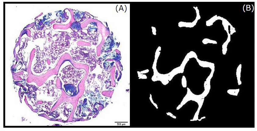
Figure 1. Microscopic image of trabecular bone (A) (pink area). The black and white picture (B) is the image modified by image segmentation technique. The white areas represent the area of trabecular bone.
RESULTS
The result of each sample from L1-L5 is reported by the image segmentation program in the form of %TBA/TA. The %TBA/TA of both male and female groups tends to decrease with increase in age. When compare the graphical representation of %TBA/TA of L1-L5 lumbar vertebrae, it is clearly seen that all lumbar vertebrae show a decrease in age in both sexes (Figures 2-4).
Table 2 shows the descriptive statistics for %TBA/TA of L1-L5 lumbar vertebrae and age for male and female groups. Comparison of the mean values of the male and female groups, shows that the %TBA/TA of L1-L5 lumbar vertebrae are not significant different. The Pearson correlation coefficients between age of each gender and %TBA/TA of L1-L5 are presented in Table 3. %TBA/TA of L2 in the male group shows the highest correlation with increase in age (r=-0.775), followed by L5, L4, L1 and L3. In the female group, the correlation of %TBA/TA of L3 is the highest (r=-0.756), followed by L1, L2, L5 and L4. When both groups are taken together the highest %TBA/TA correlation with age is L2 (r=-0.730), followed by L1, L5, L4 and L3. (Table 3).
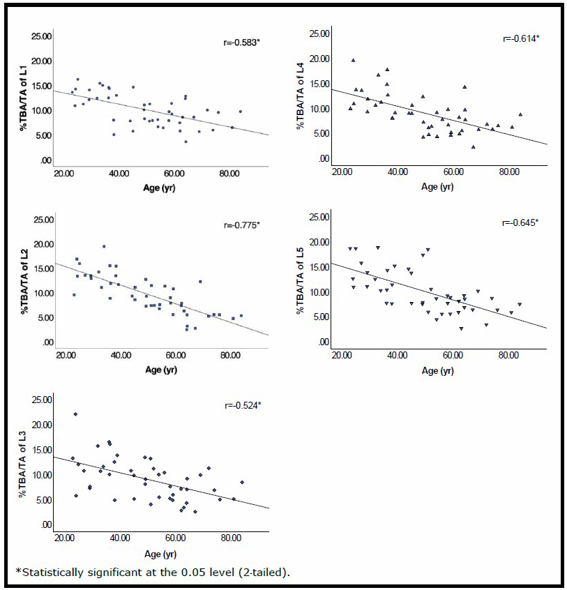
Figure 2. The %TBA/TA graph and Pearson correlation coefficients between age and L1-L5 lumbar level in the male group.
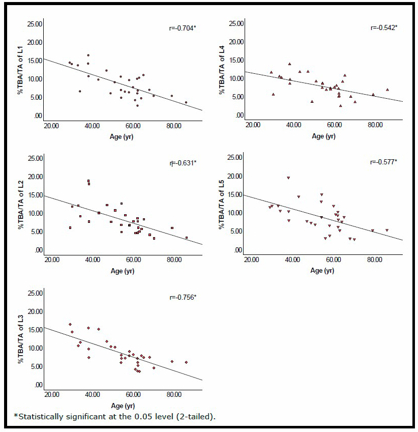
Figure 3. The %TBA/TA graph and Pearson correlation coefficients between age and L1-L5 lumbar level in the female group.
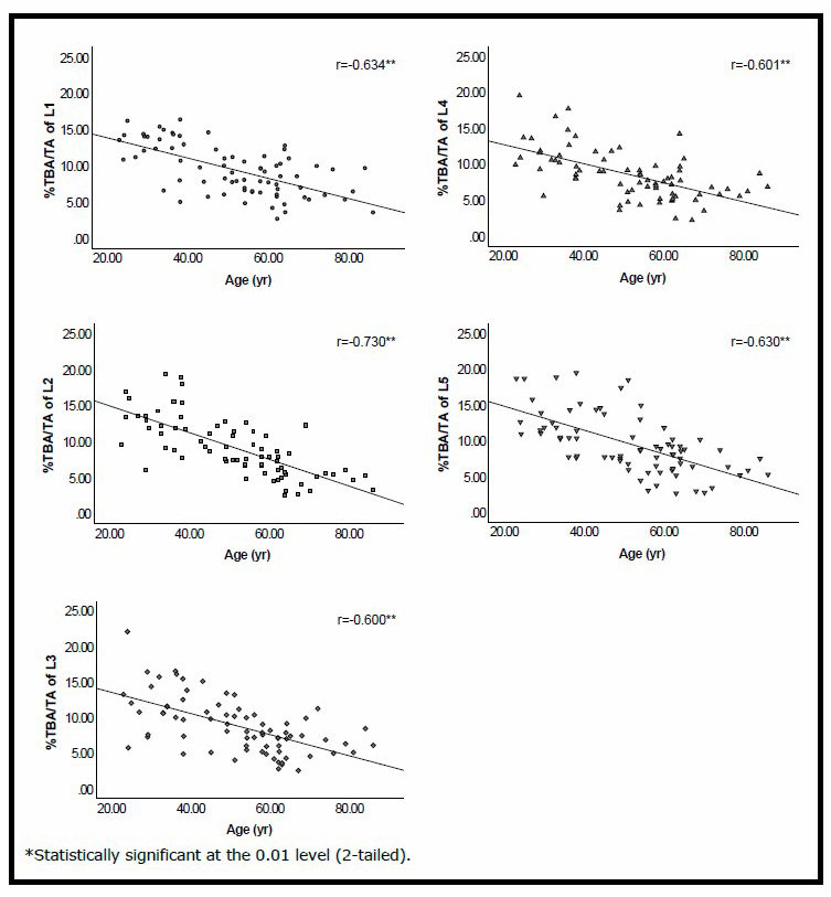
Figure 4. The %TBA/TA graph and Pearson correlation coefficients between and L1-L5 lumbar level in the mixed group.
Table 2. Descriptive statistics for age, %TBA/TA of L1-L5, and P value of the comparison between sexes.
|
Parameter |
Male (n=45) |
|
Female (n=30) |
P value |
||||||||
|
Min |
Max |
Mean |
SD |
SE |
|
Min |
Max |
Mean |
SD |
SE |
||
|
Age (yr) |
23 |
84 |
49.79 |
16.48 |
2.46 |
|
29 |
86 |
54.31 |
14.14 |
2.58 |
0.22 |
|
L1 (%) |
3.61 |
16.22 |
10.05 |
3.23 |
0.48 |
|
2.71 |
16.40 |
8.57 |
3.62 |
0.66 |
0.07 |
|
L2 (%) |
2.46 |
19.44 |
9.66 |
4.02 |
0.60 |
|
3.17 |
18.87 |
8.25 |
3.84 |
0.70 |
0.13 |
|
L3 (%) |
2.56 |
22.09 |
9.07 |
4.16 |
0.62 |
|
3.61 |
16.42 |
8.54 |
3.45 |
0.63 |
0.57 |
|
L4 (%) |
2.20 |
19.52 |
9.02 |
3.80 |
0.57 |
|
2.43 |
13.86 |
7.79 |
2.74 |
0.50 |
0.13 |
|
L5 (%) |
2.68 |
18.86 |
10.09 |
4.27 |
0.64 |
|
2.78 |
19.49 |
8.74 |
3.86 |
0.70 |
0.17 |
Note: Max: Maximum, Min: Minimum, SD: Standard Deviation, SE: Standard Error.
Table 3. Pearson correlation coefficients between age and L1-L5 of each sex.
|
Sexes |
Correlation coefficient (r) with lumbar level |
||||
|
L1 |
L2 |
L3 |
L4 |
L5 |
|
|
Male |
-0.583* |
-0.775* |
-0.524* |
-0.614* |
-0.645* |
|
Female |
-0.704* |
-0.631* |
-0.756* |
-0.542* |
-0.577* |
|
Mixed |
-0.634** |
-0.730** |
-0.600** |
-0.601** |
-0.630** |
Note: *Statistically significant at the 0.05 level (2-tailed).
**Statistically significant at the 0.01 level (2-tailed).
DISCUSSIONS
Age estimation is one of the essential pillars for preliminary individual identification in an unknown cadaver. Most age estimation methods have been performed on the pelvis and skull by morphological techniques (Rissech, et al., 2006; Miranker, 2016; Priya, 2017), however the outcome range of age is usually wide. Many studies showed that the pelvis gives the most accurate age estimation (Miranker, 2016; Priya, 2017). Singsuwan et al. (2019) studied the pelvis in a Thai population and reported that the adult acetabulum could be used as a tool for age estimation with 66% accuracy (+/–10 years) and 71% accuracy (+/–12 years) in both males and females (Singsuwan, et al., 2019). The other bone which is widely acknowledged as being a useful tool for age estimation is the skull. However, a study of suture closures in a Thai population by Ruengdit et al. (2018), using the methods of Meindl and Lovejoy (1985), Acsádi and Nemeskéri (1970), and Mann (1991), revealed poor effectiveness as regards age estimation with endocranial suture. Only obliteration of the median palatine suture, using the Mann method (1991), could be used for prediction of the age of individuals over-50-years of age (Ruengdit et al., 2018).
Even though both the pelvis and skull are useful for age estimation, unfortunately, there have been many occasions when both pelvis and skull were missing. Therefore, it is important to find some other bones to give this profile aspect of the body. It is now acknowledged that lumbar vertebrae are potentially useful in this respect. Praneatpolgrang et al. (2019) reported a high correlation between age and vertebral osteophytes in a Thai population. The correlation coefficients of male, female and mixed groups with increase in vertebral osteophytes were 0.751, 0.786 and 0.749, respectively (Praneatpolgrang, et al., 2019).
It is known that trabecular bone changes throughout human life. When age increases, the number of trabecular bones will be decreased (Chen, et al., 2013). This is true with lumbar vertebrae as well. Van Der Linden et al. reported in their study on three-dimensional simulation of age-related remodeling in trabecular bone in 2001 that bone loss increased with the number of years of remodeling (Van Der Linden, et al., 2001). There have been many more studies which have reported that the degree of trabecular bone decrease was useful for age estimation (Thomsen et al., 2002; Castillo et al., 2012; Thomsen et al., 2015).
However, since most current methods for age estimation by vertebral trabecular bones were generated from non-Thai samples they cannot be applied confidently to Thai remains, lumbar vertebrae from a Thai population were investigated in this study to assess their relationship with age. The results of this study provide the basic information to inform a full research paper to examine the accuracy of age estimation by %TBA/TA of lumbar vertebrae using this image processing technique to predict the age of unknown persons.
The number of sample size of this study was determined by the formula following Zar in 2010 (Zar, 2010). With 75 Thai cadaver samples, the power of test is no less than 80%, under the significant levels at 0.05 and the error estimation of correlation coefficient about 17%. The correlation coefficient between trabecular bone area and age is 0.89 (Castillo et al., 2012) which we used as reference value for sample size determination.
It was found in this study that histological use of H&E stain, which is routine practice in a pathological lab, can be effective with this image segmentation technique. The decrease of %TBA/TA with age is observed in both males and females (Figures 2-4) a finding which concurs with previous studies. In 2002, Thomsen et al. performed a new computerized analysis of horizontal and vertical trabeculae and found that trabecular bone number on both planes, as well as the horizontal trabecular bone thickness, decreased with age in every lumbar vertebrae (Thomsen et al., 2002). Castillo et al. (2012), reported a high correlation (r=-0.893) between age and trabecular volume of the iliac crest by using a histological technique (Castillo et al., 2012). Later, Thomsen et al. (2015) reported in their article that trabecular bone volume per total volume, connectivity density, and trabecular bone number of vertebral body and iliac crest decreased significantly with age, both in women and men (Thomsen et al., 2015).
Though %TBA/TA of males was not different from females (Table 2), the relationship between %TBA/TA of each lumbar vertebra and age was analyzed in each sex separately to see the most related-to-age lumbar level. It is evident that %TBA/TA of L2 (-0.775) and L3 (-0.756) show the highest correlation to the age of males and females, respectively (Table 3). So, if the body is male or unknown sex, L2 is suggested to be used for age estimation. But if the body is identified as a female, L3 is preferred. The correlation of L2 with age in this study is slightly lower than in a previous study by Castillo et al. in 2012 (r=-0.893) (Castillo et al., 2012). When compared to the study by Praneatpolgrang et al. the correlations between age and vertebral osteophytes and %TBA/TA are almost the same (Praneatpolgrang et al., 2019). The lumbar vertebra of highest correlation with age in this Thai male group, L2, differs from that reported in the study by Hayashi et al. (2011), which found that L3 had the lowest trabecular bone mineral density in related to increase in age (Hayashi et al., 2011).
In a case in which the body is found with the lumbar vertebra with the highest correlation missing, the next vertebra in sequence of the correlation level could be used for age estimation; specifically L5, L4, L1 and L3 respectively for male, L1, L2, L5 and L4 respectively for female and L1, L5, L4 and L3 respectively for unknown sex. However, since the correlations with age of the last two vertebrae in each group are not significantly higher, it is suggested that another method for age estimation is used in addition to this method, such as measurement of vertebral osteophytes, to increase accuracy.
CONCLUSION
In summary, this study was to find the vertebrae in each sex which give the highest degree of accuracy for age estimation by %TBA/TA based on correlation level with age. The technique of image segmentation used for %TBA/TA calculation is very convenient and cost effective as regards time. This study can be used as a pilot study which can lead to a more extensive investigation into how the %TBA/TA of lumbar vertebrae correlates to the age of Thai individuals, in which all data can be used to facilitate the process of accurate reading of the age of an unknown individual.
ACKNOWLEDGEMENT
The authors would like to thank the research administration section, Faculty of Medicine at Chiang Mai University for their financial support of this project.
CONFLICT OF INTEREST
The authors declare that they have no conflicts of interest.
REFERENCES
Albanese, J., Osley, S.E., and Tuck, A. 2016. Do group-specific equations provide the best estimates of stature? Forensic science international. 261: 154–158.
Albert, M., Mulhern, D., Torpey, M. A., and Boone, E. 2010. Age estimation using thoracic and first two lumbar vertebral ring epiphyseal union. Journal of Forensic Sciences. 55: 287–294.
Castillo, R.F., Ubelaker, D.H., and Djorojevic, M. 2012. Age estimation through histological study of trabecular volume and cortical bone width of the iliac crest. Science & Justice. 52: 177–180.
Cattaneo, C., De Angelis, D., Ruspa, M., Gibelli, D., Cameriere, R., and Grandi, M. 2008. How old am I? Age estimation in living adults: a case report. Journal of Forensic Odonto-stomatology. 26: 39–43.
Chen, H., Zhou, X., Fujita, H., Onozuka, M., and Kubo, K.-Y. 2013. Age-related changes in trabecular and cortical bone microstructure. International journal of endocrinology. 2013.
Giordano, V., Franco, J.S., Koch, H. A., Labronici, P.J., Pires, R.E., and Amaral, N.P. 2016. Age-related changes in bone architecture. The Journal of the Brazilian College of Surgeons. 43: 276–285.
Gridley, M.F. 1957. Manual of histologic and special staining technics: Armed Forces Institute of Pathology.
Hayashi, T., Chen, H., Miyamoto, K., Zhou, X., Hara, T., Yokoyama, et al. 2011. Analysis of bone mineral density distribution at trabecular bones in thoracic and lumbar vertebrae using X-ray CT images. Journal of bone and mineral metabolism. 29: 174–185.
Introna, F., and Campobasso, C.P. 2006. Biological vs legal age of living individuals. In Forensic anthropology and medicine. (pp. 57–82): Springer.
İşcan, M.Y., Loth, S.R., and Wright, R.K. 1985. Age estimation from the rib by phase analysis: white females. Journal of Forensic Science. 30: 853–863.
Martins, R., Oliveira, P.E., and Schmitt, A. 2012. Estimation of age at death from the pubic symphysis and the auricular surface of the ilium using a smoothing procedure. Forensic science international. 219: 287. e281–e287.
Miranker, M. 2016. A comparison of different age estimation methods of the adult pelvis. Journal of Forensic Sciences. 61: 1173–1179.
Mulhern, D.M., and Jones, E.B. 2005. Test of revised method of age estimation from the auricular surface of the ilium. American Journal of Physical Anthropology: The Official Publication of the American Association of Physical Anthropologists. 126: 61–65.
Pattamapaspong, N., Madla, C., Mekjaidee, K., and Namwongprom, S. 2015. Age estimation of a Thai population based on maturation of the medial clavicular epiphysis using computed tomography. Forensic science international. 246: e121–e123.
Praneatpolgrang, S., Prasitwattanaseree, S., and Mahakkanukrauh, P. 2019. Age estimation equations using vertebral osteophyte formation in a Thai population: comparison and modified osteophyte scoring method. Anatomy & cell biology. 52: 149–160.
Priya, E. 2017. Methods of skeletal age estimation used by forensic anthropologists in adults: a review. Forensic Research & Criminology International Journal. 4: 00104.
Rissech, C., Estabrook, G.F., Cunha, E., and Malgosa, A. 2006. Using the acetabulum to estimate age at death of adult males. Journal of Forensic Sciences. 51: 213–229.
Ruengdit, S., Prasitwattanaseree, S., Mekjaidee, K., Sinthubua, A., and Mahakkanukrauh, P. 2018. Age estimation approaches using cranial suture closure: A validation study on a Thai population. Journal of forensic and legal medicine. 53: 79–86.
Schmidt, C.W., and Symes, S.A. 2015. The analysis of burned human remains: Academic Press.
Singsuwan, P., Prasitwattanaseree, S., and Mahakkanukrauh, P. 2019. A study on the age estimation based on the adult acetabulum in Thai population. International Medical Journal. 26.
Thomsen, J.S., Ebbesen, E., and Mosekilde, L. 2002. Age-related differences between thinning of horizontal and vertical trabeculae in human lumbar bone as assessed by a new computerized method. Bone. 31: 136–142.
Thomsen, J.S., Jensen, M.V., Niklassen, A.S., Ebbesen, E.N., and Brüel, A. 2015. Age-related changes in vertebral and iliac crest 3D bone microstructure-differences and similarities. Osteoporosis International. 26: 219–228.
Thomsen, J.S., Niklassen, A.S., Ebbesen, E.N., and Brüel, A. 2013. Age-related changes of vertical and horizontal lumbar vertebral trabecular 3D bone microstructure is different in women and men. Bone. 57: 47–55.
Van Der Linden, J.C., Verhaar, J., and Weinans, H. 2001. A three‐dimensional simulation of age‐related remodeling in trabecular bone. Journal of Bone and Mineral Research. 16: 688–696.
Vodanović, M., Dumančić, J., Galić, I., Pavičin, I. S., Petrovečki, M., Cameriere, R., and Brkić, H. 2011. Age estimation in archaeological skeletal remains: evaluation of four non-destructive age calculation methods. Journal of Forensic Odonto-stomatology. 29: 14.
Watanabe, S., and Terazawa, K. 2006. Age estimation from the degree of osteophyte formation of vertebral columns in Japanese. Legal Medicine. 8: 156–160.
Zar JH. 2010 Biostatistical analysis Pearson Prentice–Hall. Upper Saddle River, New Jersey.
Zhang, K., Cui, J.-h., Luo, Y.-z., Fan, F., Yang, M., Li, X.-h., et al., 2016. Estimation of stature and sex from scapular measurements by three-dimensional volume-rendering technique using in Chinese. Legal Medicine. 21: 58–63.
OPEN access freely available online
Chiang Mai University Journal of Natural Sciences [ISSN 16851994]
Chiang Mai University, Thailand
https://cmuj.cmu.ac.th
Pornpath Sattarath1, Kittichai Wantanajittikul2, Sukon Prasitwattanaseree3, Jongkolnee Settakorn4, and Karnda Mekjaidee5,*
1 Forensic Osteology, Faculty of Medicine, Chiang Mai University, Chiang Mai 50200, Thailand
2 Department of Radiologic Technology, Faculty of Associated Medical Sciences, Chiang Mai University, Chiang Mai 50200, Thailand
3 Department of Statistics, Faculty of Science, Chiang Mai University, Chiang Mai 50200, Thailand
4 Department of Pathology, Faculty of Medicine, Chiang Mai University, Chiang Mai 50200, Thailand
5 Department of Forensic Medicine, Faculty of Medicine, Chiang Mai University, Chiang Mai 50200, Thailand
Corresponding author: Karnda Mekjaidee, E-mail: karnda.me@gmail.com
Total Article Views
Editor: Korakot Nganvongpanit,
Chiang Mai University, Thailand
Article history:
Received: November 10, 2020;
Revised: February 19, 2021;
Accepted: April 4, 2021;
Published online: April 30, 2021