 ISSN: 2822-0838 Online

Exploring the Sources of PM10 Burning-Season Haze in Northern Thailand Using Nuclear Analytical Techniques
Suchart Kiatwattanacharoen, Tippawan Prapamontol*, Somsorn Singharat, Somporn Chantara and Prasak ThavornyutikarnPublished Date : 2017-10-01
DOI : https://doi.org/10.12982/CMUJNS.2017.0025
Journal Issues : Number 4 ,October-December 2017
ABSTRACT
This study explored the sources of PM10 in the smoke haze during the traditional burning season in northern Thailand by determining the characteristics of the atomic elements in PM10 compared to known plant samples. The ambient air was collected from two sites (urban and peri-urban) in the Chiang Mai - Lamphun Basin. This was compared to the characteristics of the leaves from eight agricultural and forest plants predominant in the region: bamboo, grass, teak, yangna, corn, longan, lychee, and rice that were collected and burned in a combustion chamber to collect the resultant PM10. The elements – Al, Si, S, Cl, K, Ca, Ti, Cr, Mn, and Fe – were analyzed by PIXE, SEM-EDS, and μ-SXRF. Morphologies of PM10 particles were analyzed by SEM. The concentrations of the elements in the PM10 of the ambient air samples correlated highly with the PM10 from the combustion of teak, yangna, and corn leaves. The results of principal component analysis (PCA), correlations, and morphological characteristics analyzed by SEM also showed that the ambient air PM10 belonged to the same group as the PM10 from combustion of teak, yangna, and corn. A HYSPLIT trajectory model indicated that the ambient air PM10 in the Chiang Mai - Lamphun Basin was derived primarily from hotspots on the Thai-Myanmar border driven by southwest winds, as well as some hotspots in the basin itself. This study has shown that open burning of plant sources, both forest and agricultural, particularly along the Thai-Myanmar border to the southwest, is a primary source of the smoke haze in the Chiang Mai – Lamphun Basin during the dry season.
Keywords: Smoke haze, PM10, Elements, Plants, PIXE, μ-SXRF, SEM-EDS, Chiang Mai
INTRODUCTION
Air pollution is a major environmental problem in the eight provinces of upper northern Thailand. Significant open biomass burning in agricultural andforest areas, as indicated by satellite hotspot data, produces substantial smoke/haze during the dry season (February-April). During this time, the daily average concentrations of ambient air PM10 (particulate matter less than 10 μm in diameter) in the eight provinces of upper northern Thailand have exceeded Thailand’s national ambient air quality standard (120 μg/m3) for more than twenty days per year since 2007 (PCD, 2015). In addition, the maximum concentrations were 2-4 times higher than the standard and may pose a health risk to residents due to over exposure to airborne particulate-bound PAHs (Phornwisetsirikun et al., 2014; Pongpiachan et al., 2015; Naksen et al., 2017). Prior research from the analysis of some selected ions in PM10 have suggested that the sources of air pollutants during the dry season probably came from fuel combustion and agricultural activities (Chantara et al., 2012; Sillapapiromsuk et al., 2013). Wiriya et al. (2013) studied polycyclic aromatic hydrocarbon (PAHs) in PM10 and suggested that vehicle emission and biomass burning were the main sources of PM10 and PAHs in northern Thailand. Sillapapiromsuk et al. (2013) analyzed the ions in PM10 emitted by controlled burning of biomass samples in a experimental combustion chamber, including rice straw, corn residue, and leaf litter from agricultural and deciduous forest areas, using ion chromatography. These studies identified the source of PM10 by single species using a single method.
Considerable research has explored the sources of PM10 by analyzing its chemical components – such as PAHs, ions, and elements. The United States Environmental Protection Agency (US EPA) recommends analyzing 16 priority compounds in airborne particulate-bound PAHs, including naphthalene, acenaphthylene, acenaphthene, fluorene, phenanthrene, anthracene, fluoranthene, pyrene, benzo[a]anthracene, chrysene, benzo[b]fluoroanthene, benzo[k]fluoroanthene, benzo[a]pyrene, dibenzo[a,h]anthracene, benzo[ghi]perylene, and indeno[1,2,3-cd] pyrene. These have been commonly analyzed by gas chromatography coupled with mass spectrometry (GC–MS) (Liu et al., 2007; Wiriya et al., 2013; Bian et al., 2016) and high performance liquid chromatography with UV-vis detection (HPLC-UV) (Wu and Chang, 1997; Bacaloni et al., 2004). A study of ambient air at heavily trafficked roads and in urban residential zones in Bangkok, Thailand used multivariate statistics to investigate the influence of anthropogenic activities on variations in the contents of 18 metals; the average concentrations of PM10-bounded metals (Al, V, Cr, Mn, and Fe) were similar to those of other urban cities around the world (Pongpiachan and Ilijima, 2016).
Highly toxic elements, such as As, Pb, Cd, Hg, Zn, Ni, Cu, Cr, and others, such as Fe, Ca, Ba, and Mn, which are derived from the earth’s crust or re-suspended soil (Gonzalez-Rodriguez et al., 2005), have been analyzed using inductively coupled plasma-optical emission spectroscopy (ICP-OES), atomic emission spectroscopy (ICP-AES), and mass spectrometry (ICP-MS) (Ochsenkuhn-Petropoulou and Ochsenkuhn, 2001; Gonzalez-Rodriguez et al., 2005; Karar et al., 2006; Kulshrestha et al., 2009; Flores-Rangel et al., 2015); atomic absorption spectroscopy (AAS) (Kulshrestha et al., 2009); particle-induced x-ray emission (PIXE) (Ferreira da Silva et al., 2010); synchrotron radiation micro X-ray fluorescence spectroscopy (μ-SXRF) (López et al., 2011; Pongpiachan et al., 2012a, 2012b); scanning electron microscopy-energy dispersive spectroscopy (SEM-EDS) (Flores-Rangel et al., 2015); anodic stripping voltammetry (ASV) (Ochsenkuhn-Petropoulou and Ochsenkuhn, 2001); and instrumental neutron-activation analysis (INAA) (Ochsenkuhn-Petropoulou and Ochsenkuhn, 2001). Some ions, such as sulfate (SO4 2-), nitrate (NO3-), ammonium (NH4+), sodium (Na+), calcium (Ca2+), potassium (K+), magnesium (Mg2+), bicarbonate (HCO3-), and chloride (Cl-), have been analyzed by AAS and ion chromatography (Sillapapiromsuk et al., 2013; Zhamsueva et al., 2015).
The sources of PM10 in upper northern Thailand, especially in the Chiang Mai - Lamphun Basin, its most populated center, have not yet been clearly identified. To determine more precisely the burning sources, both material and location, further analytical techniques are required. Thus, this study explored the sources of PM10 in the smoke haze during the traditional burning season in northern Thailand by determining the characteristics of the atomic elements in PM10 compared to the combustion of known plant samples using PIXE, SEMEDS, and μ-SXRF analyses.
MATERIALS AND METHODS
Collection and preparation of leaf samples
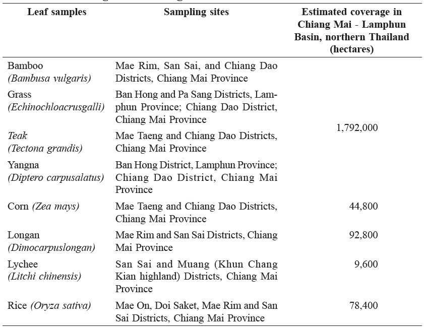
The four most common agricultural crops in upper northern Thailand are rice (591,360 hectares), corn (383,790 hectares), longan (133,900 hectares), and lychee (19,440 hectares); together they account for 1.1 m hectares, or approximately two-thirds of the total agricultural land (1.7 m hectares). The total forest area in upper northern Thailand is about 5.7 m hectares, or more than three times the agricultural area; bamboo, grass, teak, and yangna are common in the forests (Agricultural Statistics of Thailand, 2014). The leaves from these eight common agricultural and forest plants (bamboo, grass, teak, yangna, corn, longan, lychee, and rice) were collected from nine sites (Table 1) in the Chiang Mai - Lamphun Basin (Figure 1) during October and November, 2010. One kilogram of each leaf was collected and equally divided into two bags. The samples were stored at room temperature under controlled humidity using silica gel until the relative humidity levels dropped below 10%.
Table 1. Leaf samples collected from the Chiang Mai - Lamphun Basin with their estimated ground coverage.
Controlled combustion of leaf samples
The leaf samples collected from each sampling site were separately burnt in a controlled combustion chamber according to Sillapapiromsuk’s method (Sillapapiromsuk et al., 2013). Five grams of dried-leaf samples were placed in the combustion chamber and burnt for about 1 minute using LPG gas. PM10 was collected from air in the chamber for about 66 minutes, until the air pressure gauge showed zero bar. PM10 was completely released from 1 m3 of air volume of the combustion chamber at an airflow rate of 15 liters per minute. When the combustion was complete, a valve between the combustion and storage chambers was opened to allow the air in the combustion chamber to flow to the storage chamber, which was connected to a GENT Air Sampler, IAEA standard, with a SFU stacked filter unit (Gent, Belgium). A polycarbonate, thin, nuclear pore film filter with a diameter of 47 mm and pore size of 8 μm (Whatman, U.S.A.) was used to collect PM10. The filter was pre-weighed before collecting PM10. Before combustion of each sample, the chamber was cleaned by evacuation.
The filters with PM10 were stored in a desiccator to eliminate moisture for 24 hours. The filter was placed in a plastic package with plastic clamp and placed on defect-free paper; information (sample ID, sampling site, date of collection, and pre- and post-weight mass) were recorded on the label attached to the top cover of each package.
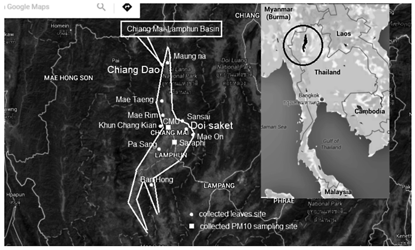
Figure 1. Map of leaf sampling sites in the Chiang Mai - Lamphun Basin and ambient air PM10 sampling sites. Site 1: Chiang Mai University (urban) (CMU, 18° 47ˊ N, 98° 57ˊ E); Site 2: Watnantaram School (peri-urban), Saraphi District (18° 44ˊ N, 99° 2ˊ E), about 20 km south of Chiang Mai City.
Note: Map modified from Google Map, 1 May 2016.
Collection and preparation of ambient air PM10
Ambient air PM10 samples were collected from two sites in the Chiang Mai - Lamphun Basin during the smoke-haze season. Site 1 (urban) was located at the Research Institute for Health Sciences (RIHES) on the main campus of Chiang Mai University (CMU) (18° 47ˊ N, 98° 57ˊ E). Site 2 (peri-urban) was at the Watnantaram School (WT) (18° 44ˊ N, 99° 2ˊ E) in Saraphi District, Lamphun (Figure 1). Ambient air PM10 samples were collected using a GENT air sampler, the same instrument used for leaf combustion. The air intake was 1.5 m above the ground. PM10 was collected at an airflow rate of 15 liters per minute, as described in the previous section. Twenty-four hour samples were collected every other day at 9:00 a.m. at both sampling sites from March to May 2010.
Backward-trajectory analysis
To investigate the location of the source that was responsible for the long range transport of PM10, 24-hour backward trajectories of air masses arriving at CMU (Site 1) were determined using Hybrid Single-Particle Langrangian Integrated Trajectory model version 4 (HYSPLIT4) during 10-13 March, 2010, the period with the highest levels of ambient air PM10 that year (PCD, 2010). Trajectories were calculated at heights of 100, 500, and 1,000 m above ground level at hourly intervals.
PIXE technique analysis
The elements in the PM10 samples (n=64) from leaf combustion and ambient air were analyzed, both qualitatively and quantitatively, by PIXE technique using a
tandem accelerator (ion-beam analysis) at the Plasma and Beam Physics Research Facility, Department of Physics and Material Sciences, Chiang Mai University, Thailand. To prepare samples for PIXE, the ionization chamber was vacuumed for about 90 minutes at 1.2x10⁻⁵ to 1.2x10⁻⁶ bar. Then, photon energy (2 MeV) was applied to each sample for 6 minutes. A device made from a 10x10x2 cm aluminum rod drilled with nine holes (1.2 cm wide and 1 cm deep) held the plastic grip ring for holding the sample filter. The data of the elemental components (Al, Si, S, Cl, K, Ca, Ti, Cr, Mn, and Fe) were obtained. The concentration of each sample in the small area hit by the high-energy proton beam (in ng/cm2) was calculated as equivalent to the entire filter area, and a conversion formula was used to calculate the element mass of particles in 1 m3 of collected air.
SEM-EDS analysis
SEM-EDS was applied to characterize the PM10 morphology and elemental composition. PM10 samples (n=45, as some samples failed during analysis) were analyzed by SEM-EDS (FEI model Quanta 200 3D EM, U.S.A.) at the Science and Technology Service Center, Faculty of Science, Chiang Mai University, Thailand. SEM-EDS was calibrated using reference standard no. 5936. Pieces of thin film sample were mounted and stuck on a brass rod. Samples in the holder were filled and locked using non-metallic adhesive tape. Then, the film sample was coated to a solid form with gold by vacuum-coating with a Gold Sputter Coater. The fine coating of gold made the samples electrically conductive. The sample holder was installed in the analyzing chamber in a vacuum (1x10⁻⁴ mbar), and then analyzed at 15kV of accelerating voltage with the current set at 0.14 nA. A Si (Li) detector was placed 10 mm away from the sample to be analyzed. The X-ray detection limit was nearly 0.1%, with the SEM-EDS system at 133 eV resolution. Magnification of 5,000 times allowed for easily locating the center of the X-ray for exposure in the SEM-EDS mode, and for a well-focused image. Elemental composition of the particles was identified with SEM-EDS spectra.
μ-SXRF analysis
Elements in the PM10 samples (n=32, as some samples failed during analysis) were analyzed by μ-SXRF at the Siam Synchrotron Research Institute (SLRI, Nakhon Ratchasima, Thailand). Calibration was performed with Zn energy (9659keV). The sample filter was placed on a sample holder that was installed in a high-vacuum analytical chamber; Ar gas was filled to 93.6 mbar and He gas was filled until pressure of 1 ATM (990 mbar). The characteristic X-ray emitted after synchrotron light hit the sample filter was 100 mm. A multichannel analyzer (MCA) was used to obtain the elemental distribution on the peak energy spectrum. The peak energy of each characteristic X-ray was unique for each element. As μ-SXRF spectroscopy measures the energy of the outgoing radiation, and the energy of fluorescent radiation is element-specific, the amount of each element in a sample can be determined.
Statistical analysis
Pearson’s correlation coefficients (r) were calculated using concentrations of the elements in PM10 analyzed by PIXE and SEM-EDS. Principal component analysis (PCA) was performed using all the element concentrations in PM10 from leaf combustion and ambient air obtained at the CMU and Saraphi sites.
RESULTS
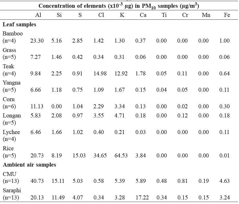
Elements were analyzed by PIXE and SEM-EDS. The concentrations of elements obtained by PIXE analysis in PM10 from leaf combustion and ambient air are shown in Table 2. Al, S, Cl, K, Ca, and Fe were found in all the samples. The concentrations of Al, Si, Cl, K, and Ca in PM10 derived from the combustion of rice and teak leaves were relatively high.
Table 2. Concentration of elements in PM10 samples from PIXE analysis.
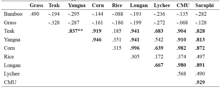
The concentrations of elements in PM10 from leaf combustion and ambient air showed generally high correlations, as shown in Table 3. High correlation coefficients were obtained among bamboo, corn, lychee, teak, longan, yangna, and ambient air PM10 derived from the urban site (CMU), and among bamboo, lychee, teak, yangna, and ambient air PM10 from the peri-urban site (Saraphi).
Table 3. Correlation coefficients of elements from PIXE analysis to PM10 ratio among PM10 samples from leaf combustion and ambient air.
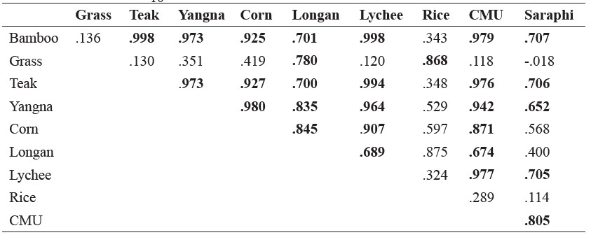
Note: Bold represents significant correlation at p<0.05.
The concentrations of elements obtained by SEM-EDS in PM10 from leaf combustion are shown in Table 4. In the leaf samples, the concentration of Al was relatively high in grass and longan; Si only in corn; S in bamboo, grass, teak, and rice; Cl in yangna, corn, longan, and lychee; K in teak, yangna, corn, and lychee; and Mn only in longan. In the ambient PM10 samples from both collection sites, Al and Mn were relatively higher than the other elements.
Table 4. Ratio of elements per PM10 of leaf samples by SEM-EDS.
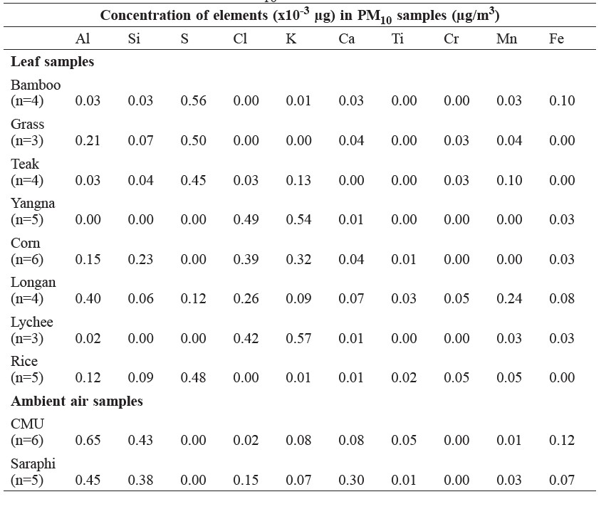
Table 5. Correlation coefficients of elements from SEM-EDS analysis to PM10 ratio among PM10 samples from leaf combustion and ambient air.
Note: Bold represents significant correlation at p<0.05.
Correlation coefficients among the concentrations of elements in the PM10 samples from leaf combustion and ambient air analyzed by SEM-EDS are shown in Table 5. Teak, yangna, corn, and longan were highly correlated with PM10 derived from both collection sites.
Morphologies and elemental compositions of PM10 analyzed by SEM-EDS
PM10 from rice leaf combustion were particles of large grain size of about 10-20 μm with a non-uniform and irregular shape (Figure 2a). Some KCl crystals (square shape) were found in visual images. PM10 from bamboo leaf combustion were particles of large grain size of about 20-30μm, irregular shape, and porous surface (Figure 2b). PM10 from grass combustion were particles of large grain size of about 30-40 μm with smooth, porous surface and some tiny cilia (Figure 2c). PM10 from corn leaf combustion were particles of moderate grain size of less than 5μm with an irregular shaped, non-porous surface microstructure (Figure 2d). PM10 from longan leaf combustion were particles of large grain size of more than 10μm with an irregular-shape and a non-porous surface (Figure 2e). PM10 from lychee leaf combustion were particles of size less than 1μm with spherical shape (Figure 2f). PM10 from teak leaf combustion were particles of moderate grain size of 1-5μm with irregular shape and non-porous surface (Figure 2g). PM10 from yangna leaf combustion were particles of small grain size of less than 1μm with irregular shape (Figure 2h). The ambient air PM10 samples collected at the urban (CMU) site were particles of fine grain size, including small and moderate sized particles with irregular shape and non-porous surface (Figure 2i). The ambient air PM10 samples collected at the peri-urban (Saraphi) site were particles of small grain size at less than 1μm with irregularly shape and non-porous surface without cilia structure (Figure 2j).
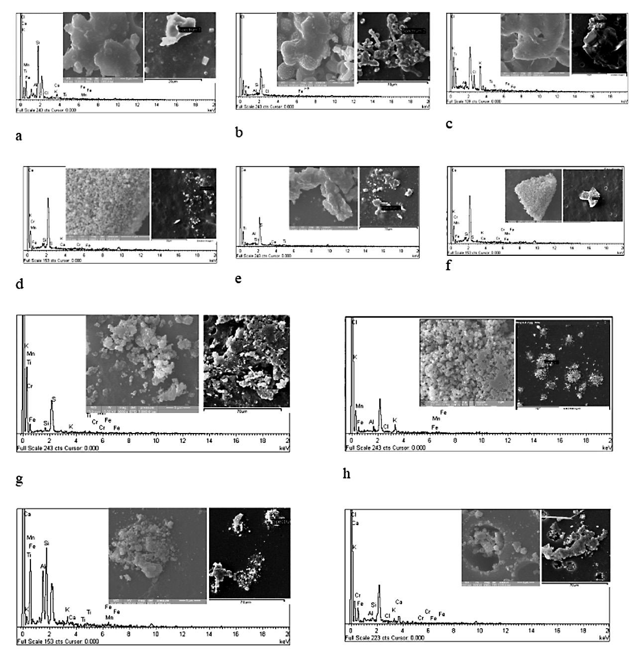
Figure 2. PM10 morphology of elements of burned leaf and ambient air samples: (a) rice, (b) bamboo, c) grass, (d) corn, (e) longan, (f) lychee, (g) teak, (h) yangna, (i) CMU ambient air samples, and (j) Saraphi ambient air samples.
Table 6. Sample types in each group categorized by their morphologies derived from SEM-EDS.
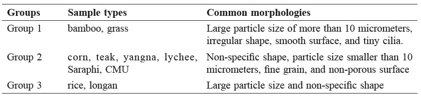
The PM10 particles shown in Figure 2 were classified into three groups by their morphological characteristics, such as shape, surface (smooth, irregular, or porous), the presence of tiny cilia, and grain size (small, moderate, and large). The results are shown in Table 6. PM10 from bamboo and grass combustion belonged to Group 1, which were particles of large grain size of more than 10μm with irregular shape, smooth surface, and tiny cilia. PM10 from corn, teak, yangna, and lychee combustion and ambient air belonged to Group 2, which were particles smaller than 10μm with non-specific shape. PM10 from rice and longan belonged to Group 3, which were large particles with non-specific shape.
Table 7. Elements found in PM10 samples of leaf combustion and ambient air from PIXE, SEM-EDS, and μ-SXRF.
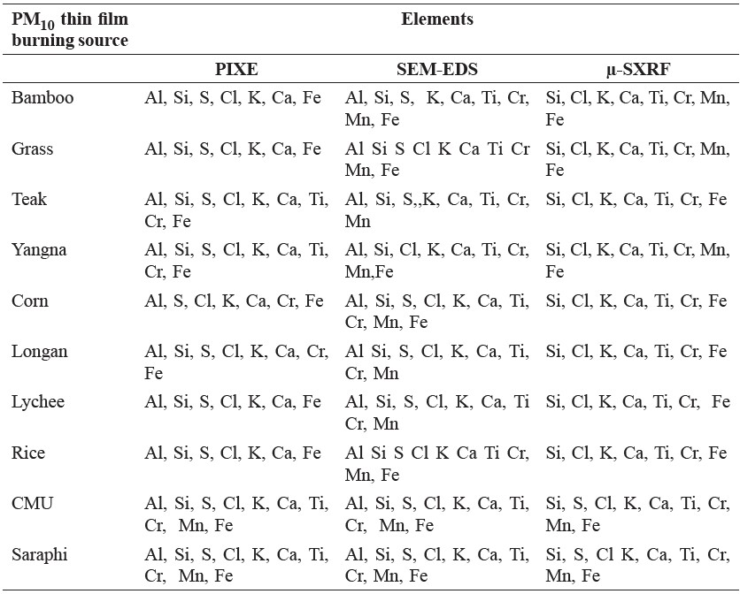
PIXE, SEM-EDS, and μ-SXRF techniques are able to detect similar elements, including the Al, Si, S, Cl, K, Ca, Ti, Cr, Mn, and Fe found in the eight plant samples and two ambient samples (Table 7).
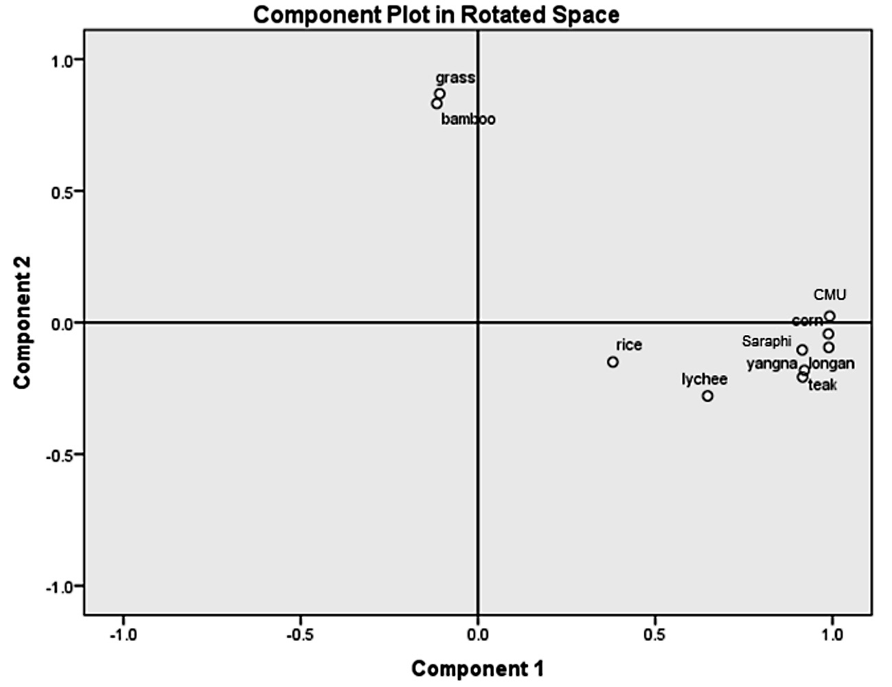
Figure 3. PCA of elements in PM10 analyzed by SEM-EDS in samples from leaf combustion and ambient air from the CMU and Saraphi sites.
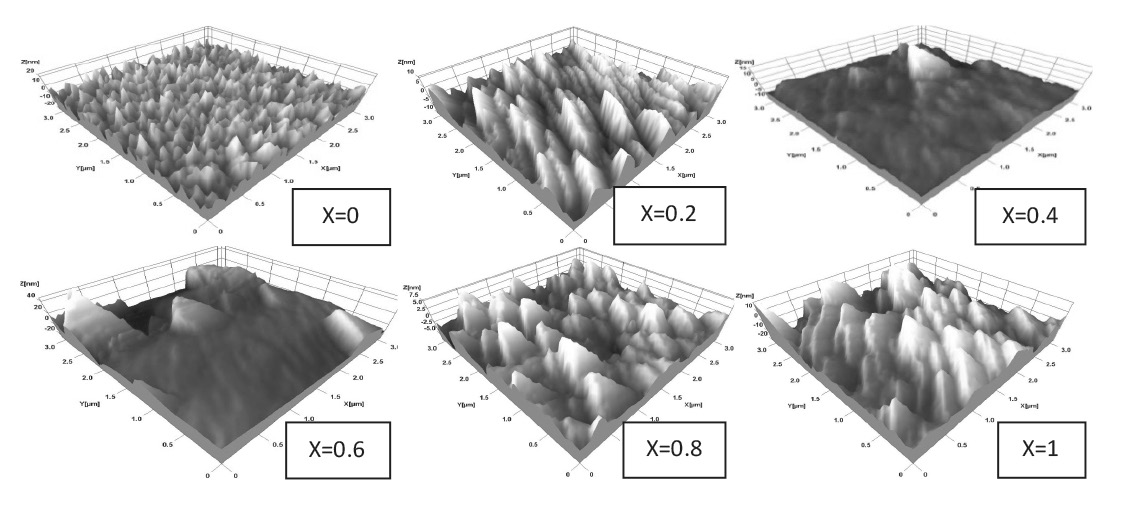
Figure 4. PCA of elements in PM10 analyzed by PIXE in samples from leaf combustion and ambient air from the CMU and Saraphi sites.
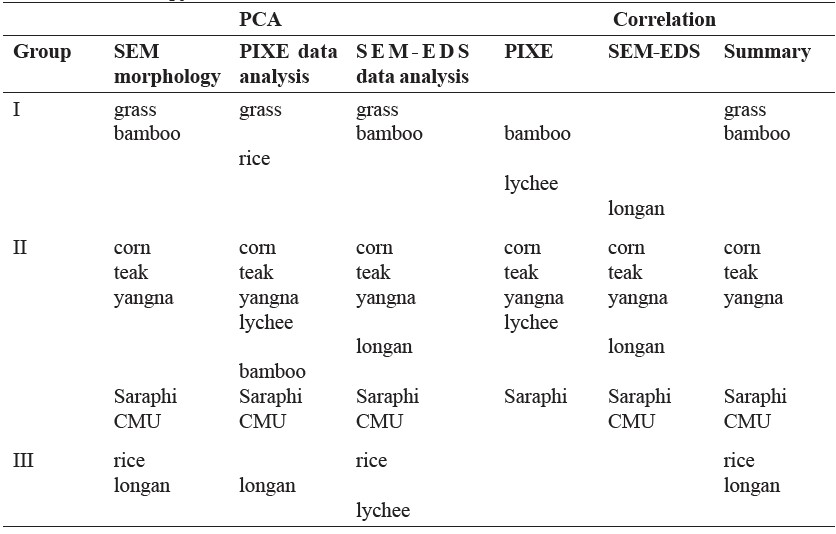
The elements analyzed by SEM-EDS in PM10 from the leaf combustion and ambient air samples were subjected to principal component analysis (Figure 3). PCA analysis was also performed using data from the PIXE analysis (Figure 4). The result differed from that of SEM-EDS. Table 8 summarizes the groups classified by SEM morphology, PCA, and correlation analysis. Although some kinds of leaves were classified to different groups by different analyses, the following were consistently classified: bamboo and grass (Group I); corn, teak, and yangna (Group II); and longan and rice (Group III). Ambient air PM10 samples from the urban (CMU) and peri-urban (Saraphi) sites were classified as Group II.
Table 8. Data as grouped by the various analytical methods. SEM morphology was group by physical visibility; PCA statistical analysis using data from PIXE and SEM-EDS. The correlations used the ratio of elements in PM10 of each leaf and ambient air sample.
HYSPLIT trajectory model
To identify the possible origins of the PM10 in the Chiang Mai - Lamphun Basin, 24-h backward trajectories during February to March 2010 were performed by HYSPLIT trajectory model.
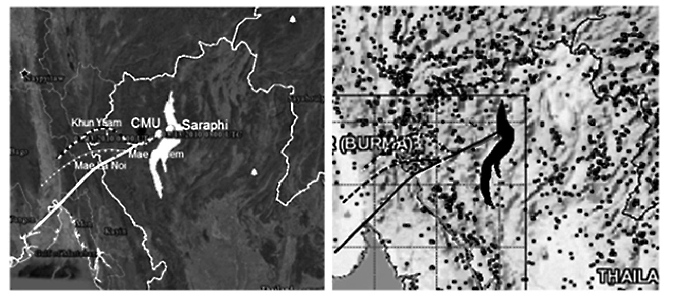
Figure 5. (a) Backward trajectories associated with smoke/haze arriving at the CMU site on 13 March 2010 and (b) hotspots in northern Thailand and the surrounding region during 10-13 March 2010.
From the HYSPLIT analysis, the winds at 100, 500, and 1,000 m above grond level blew from the southwest of the Chiang Mai - Lamphun Basin, as shown in Figure 5(a), generally from the direction of Mae Hong Son Province and bordering areas of Myanmar. This wind direction was coincident with a dense area of hotspots in the Mae Hong Son Province – Myanmar border area, as shown in Figure 5(b). This area includes a high density of cornfields (55,166 hectares) within a forest area of about 235,200 hectares (Official Statistics Thailand, 2014). Overall, about 88% of Mae Hong Son Province is forest. In upper northern Thailand, forest fires and agricultural biomass burning frequently occur during the dry and postharvest season of January to April.
DISCUSSION
Our findings suggested that sources of PM10 at CMU and Saraphi sites in the haze produced during the biomass-burning season were from the burning of teak, yangna, and corn (Table 8). These three out of eight common plants cover a significant portion of the growing area in the Chiang Mai - Lamphun Basin, northern Thailand (Table 1).
The morphology of PM10 collected from the CMU and Saraphi sites matched closely with the morphology of PM10 from corn, teak, yangna, and lychee (Table 6). Statistical analyses (PCA and correlation methods) also played an important role in identifying these sources of burning. PCA of element concentrations of PM10 analyzed by SEM-EDS technique indicated four plants: corn, teak, yangna, and longan (Figure 3). PCA of element concentrations of PM10 analyzed by PIXE technique found five plants: corn, teak, yangna, lychee, and bamboo (Figure 4). Correlation analysis of the element concentrations analyzed from both the SEMEDS and PIXE techniques found overlapping plant types. With PIXE technique, the element concentrations from the CMU site and bamboo, teak, yangna, corn, longan, and lychee were highly correlated; and the Saraphi site and bamboo, teak, yangna, and lychee were highly correlated (Table 3). With SEM-EDS technique, the element concentrations from the CMU site and teak, yangna, corn, and longan were highly correlated; and the Saraphi site and teak, yangna, corn, and longan were highly correlated (Table 5). μ-SXRF technique confirmed the elements found in the PM10 from PIXE and SEM-EDS (Table 7). Furthermore, element concentrations in PM10 from CMU and Saraphi sites analyzed by both PIXE and SEM-EDS techniques were highly correlated (Tables 3 and 5). In summary, as shown in Table 8, elemental concentrations analyzed by the different analytical techniques and accompany statistical methods pointed to three plant-burning types, i.e., corn, teak, and yangna, that related to airborne PM10 at the CMU and Saraphi sites.
The wind bringing PM10 to the CMU site came from the southwest of the Chiang Mai - Lamphun Basin during the study period (Figure 5a); this was coincident with dense hotspot areas in Mae Chaem District (Chiang Mai Province), Mae Hong Son Province, and the Myanmar border area (Figure 5b).
Several related, but different, biomass-burning studies in northern Thailand have been reported, including: analysis of the elements in airborne particles using ICP-OES (Khamkaew et al., 2016); chemical tracers, i.e., potassium ion and levoglucosan as an indicator of biomass-burning activities (Lin et al., 2014); PM10 and its ion composition emitted from biomass burning in a combustion chamber for estimating open-burning emissions (Sillapapiromsuk et al., 2013); concentrations of airborne PM10 and PM10-bound PAHs for source identification (Wiriya et al., 2013).
Several studies of PM10 sources in northern Thailand have been reported (Kim Oanh et al., 2011; Phoothiwut and Junyapoon, 2013), but they have focused on the rice straw burning season that occurs in December/January after the rice harvest every year, in contrast to the smoke/haze during the dry season burning in February to March studied here. To the best of our knowledge, our study is the first of its kind to identify the types of plants that were burnt and contributed to airborne PM10 in the Chiang Mai - Lamphun Basin during the smoke/haze season.
This study is also among only a few to employ nuclear analytical techniques to determine the elements in PM10. Although they require costly instrumentation, their non-destructive analytical characteristics enable repetition in measurement (Yatkin et al., 2016; Turner et al., 2017) and allow for analysis by multiple techniques. We were able to use the filter samples (47 mm diameter) we collected with three different techniques – PIXE (n=64), SEM-EDS (n=45), and μ-SXRF (n=32) – since each technique required a minute sample size only (Huang et al., 2012). Furthermore, data from the same samples using the three different techniques strongly enhanced the statistical analysis used to identify the source of the PM10 particles.
The advantage of this study was its application of three different nuclear analytical techniques to analyze the elemental composition of airborne particles (PM10) and two statistical methods to analyze the their correlation. This identified corn, teak, and yangna as probable burning sources that contributed to the PM10 in the Chiang Mai - Lamphun Basin during the annual smoke/haze burning season in northern Thailand (Table 8). However, this studies findings may be limited by the small number of plant types investigated. Although the eight plant types studied here account for a large portion of the plants in the Chiang Mai - Lamphun Basin, the backward trajectory model showed that the burning sources probably originated outside and far beyond the basin, i.e., Mae Hong Son Province and the Myanmar border areas. More plant types including deciduous plants and other agricultural crop plants warrant further investigation.
ACKNOWLEDGEMENTS
Chiang Mai University supported this study. We are also indebted for the support from the Commission for Higher Education through the National Research University Program, Chiang Mai University. This work could not have been completed without the facility support of the Research Institute of Health Sciences, Chiang Mai University; the Department of Physics and Materials, Chiang Mai University; and the SIAM Synchrotron Light Research Institute, SLRI, Muang District, Nakhon Ratchasima Province, Thailand.
REFERENCES
Agricultural Statistics of Thailand. 2014. Office of Agricultural Economics, Ministry of Agriculture and Cooperatives Bangkok, Thailand.
Bacaloni, A., Cafaro, C., de Giorgi, L., Ruocco, R., and Zoccolillo, L. 2004. Improved Analysis of Polycyclic Aromatic Hydrocarbons in Atmospheric Particulate Matter by HPLC-Fluorescence. Annali di Chimica. 94: 751-759. https://doi.org/10.1002/adic.200490093
Bian, Q., Alharbi, B., Collett, J., Jr, Kreidenweis, S., and Pasha, M.J. 2016. Measurements and source apportionment of particle-associated polycyclic aromatic hydrocarbons in ambient air in Riyadh, Saudi Arabia. Atmospheric Environment. 137: 186-198. https://doi.org/10.1016/j.atmosenv.2016.04.025
Chantara, S., Sillapapiromsuk, S., and Wiriya, W. 2012. Atmospheric pollutants in Chiang Mai (Thailand) over a five-year period (2005-2009), their possible sources and relation to air mass movement. Atmospheric Environment. 60: 88-98. https://doi.org/10.1016/j.atmosenv.2012.06.044
Ferreira da Silva, M., Vicente de Assuncao, J., de Fatima Andrade, M., and Pesquero, C.R. 2010. Characterization of metal and elements contents of particulate matter (PM10) emitted by vehicles running on Brazilian fuels-hydrated ethanol and gasoline with 22% of anhydrous ethanol. Journal of Toxicology and Environmental Health, Part A. 73: 901-909. https://doi.org/10.1080/15287391003744849
Flores-Rangel, R.M., Rodríguez-Espinosa, P.F., de Oca-Valero, J.A.M., Mugica-Álvarez, V., Ortiz-Romero-Vargas, M.E, Navarrete-López, M., Dorantes-Rosales, H.J, and Morales-García, S.S. 2015. Temporal variation of PM10 and metal concentrations in Tampico, Mexico. Air Quality, Atmosphere & Health. 8: 367-378. https://doi.org/10.1007/s11869-014-0291-6
Gonzalez-Rodriguez, M.J., Arrebola Liebanas, F.J., Garrido Frenich, A., Martinez Vidal, J.L, and Lopez, S. 2005. Determination of pesticides and some metabolites in different kinds of milk by solid-phase microextraction and low-pressure gas chromatography-tandem mass spectrometry. Analytical and Bioanalytical Chemistry. 382: 164-172. https://doi.org/10.1007/s00216-005-3144-1
Huang, D., Xiao, C., Ni, B., Tian, W., Zhang, Y., Wang, P., Liu, C., Zhang, G., Sun, H., Zhang, H., and Zhao, C. 2012. Study on homogeneity of multielements for a stream sediment matrix material with nuclear analytical techniques. Journal of Radioanalytical and Nuclear Chemistry. 291: 573-577. https://doi.org/10.1007/ s10967-011-1330-5
Karar, K., Gupta, A.K., Kumar, A., and Biswas, A.K. 2006. Characterization and identification of the sources of Chromium, Zinc, Lead, Cadmium, Nickel, Manganese and Iron in PM10 particulates at the two sites of Kolkata, India. Environmental Monitoring and Assessment. 120: 347-360. https://doi.org/ 10.1007/s10661-005-9067-7
Khamkaew, C., Chantara, S., Janta, R., Pani, S.K., Prapamontol, T., Kawichai, S., Wiriya, W., and Lin, N.H. 2016. Investigation of biomass burning chemical components over Northern Southeast Asia during 7-SEAS/BASELInE 2014 campaign. Aerosol and Air Quality Research. 16: 2655-2670. https://doi.org/ 10.4209/aaqr.2016.03.0105
Kim Oanh, N.T., Ly, B.T., Tipayarom, D., Manandhar, B.R., Prapat, P., Simpson, C.D., and Sally Liu, L.J. 2011. Characterization of particulate matter emission from open burning of rice straw. Atmospheric Environment. 45: 493-502. https://doi.org/10.1016/j.atmosenv.2010.09.023
Kulshrestha, A., Satsangi, P.G., Masih, J., and Taneja, A. 2009. Metal concentration of PM2.5 and PM10 particles and seasonal variations in urban and rural environment of Agra, India. Science of the Total Environment. 407: 6196-6204. https://doi.org/10.1016/j.scitotenv.2009.08.050
Lin, N.H., Sayer, A.M., Wang, S.H., Loftus, A.M., Hsiao, T.C., Sheu, G.R., Hsu, N.C., Tsay, S.C., and Chantara, S. 2014. Interactions between biomass-burning aerosols and clouds over Southeast Asia: Current status, challenges, and perspectives. Environmental Pollution. 195: 292-307. https://doi.org/10.1016/ j.envpol.2014.06.036
Liu L.B., Hashi Y., Liu M., Wei Y., and Lin J.M. 2007. Determination of particle-associated polycyclic aromatic hydrocarbons in urban air of Beijing by GC/MS. Analytical Sciences. 23: 667-671. https://doi.org/10.2116/analsci.23.667
López, M.L., Ceppi, S., Palancar, G.G., Olcese, L.E., Tirao, G., and Toselli, B.M. 2011. Elemental concentration and source identification of PM10 and PM2.5 by SR-XRF in Córdoba City, Argentina. Atmospheric Environment. 45: 5450-5457. https://doi.org/10.1016/j.atmosenv.2011.07.003
Naksen, W., Kawichai, S., Srinual, N., Salrasee, W., and Prapamontol, T. 2017. First evidence of high urinary l-hydroxypyrene level among rural school children during smoke haze episode in Chiang Mai province, Thailand. Atmospheric Pollution Research. 8: 418-427. https://doi.org/10.1016/j.apr.2016.11.002
Ochsenkuhn-Petropoulou, M., and Ochsenkuhn K.M. 2001. Comparison of inductively coupled plasma-atomic emission spectrometry, anodic stripping voltammetry and instrumental neutron-activation analysis for the determination of heavy metals in airborne particulate matter. Fresenius’ Journal of Analytical Chemistry. 369: 629-632. https://doi.org/10.1007/s002160100769
Official Statistics Thailand. 2014. Corn planted area by province in Thailand.
PCD. 2010. Thailand state of pollution report 2010. Pollution Control Department.
PCD. 2015. Manual report: Air quality data monitoring. Pollution Control Department. http://aqmthai.com/
Phoothiwut, S., and Junyapoon, S. 2013. Size distribution of atmospheric particulates and particulate-bound polycyclic aromatic hydrocarbons and characteristics of PAHs during haze period in Lampang Province, Northern Thailand. Air Quality, Atmosphere and Health. 6: 397-405. https://doi.org/ 10.1007/s11869-012-0194-3
Phornwisetsirikun, W., Prapamontol, T., Rangkakulnuwat, S., Chantara, S., Tavornyutikarn, P. 2014. Elevated ambirent PM10 levels affecting respiratory health of schoolchildren in Chiang Mai, Thailand. Chiang Mai University Journal of Natural Sciences. 13(3): 345-354. https://doi.org/10.12982/CMUJNS.2014.0040
Pongpiachan, S., and Iijima, A. 2016. Assessment of selected metals in the ambientair PM10 in urban sites of Bangkok (Thailand). Environmental Science and Pollution Research International. 23: 2948-2961. https://doi.org/10.1007/s11356-015-5877-5
Pongpiachan, S., Thumanu, K., Kositanont, C., Schwarzer, K., Prietzel, J., Hirunyatrakul, P., and Kittikoon, I. 2012a. Parameters influencing sulfur speciation in environmental samples using sulfur K-Edge X-Ray Absorption Near-Edge Structure (XANES). Journal of Analytical Methods in Chemistry. Article ID 659858, 12 pages. http://www.hindawi.com/journals/jamc/aip/659858/.http://dx.doi.org/10.1155/2012/659858
Pongpiachan, S., Thumanu, K., Na Pattalung, W., Hirunyatrakul, P., Kittikoon, I., Ho, F.K., and Cao, J.J. 2012b. Diurnal variation and spatial distribution effects of Sulfur Speciation in Aerosol Samples as Assessed by X-ray Absorption Near-edge Structure (XANES). Journal of Analytical Methods in Chemistry. Article ID 696080, 10 pages. http://www.hindawi.com/journals/jamc/aip/696080/. http://dx.doi.org/10.1155/2012/696080.
Pongpiachan, S., Tipmanee, D., Khumsup, C., Kittikoon, I., and Hirunyatrakul, P. 2015. Assessing risks to adults and preschool children posed by PM2.5-bound polycyclic aromatic hydrocarbons (PAHs) during a biomass burning episode in Northern Thailand. Science of the Total Environment. 508: 435-444. http://dx.doi.org/10.1016/j.scitotenv.2014.12.019
Sillapapiromsuk, S., Chantara, S., Tengjaroenkul, U., Prasitwattanaseree, S., and Prapamontol, T. 2013. Determination of PM10 and its ion composition emitted from biomass burning in the chamber for estimation of open burning emissions. Chemosphere. 93: 1912-1919. http://dx.doi.org/10.1016/ j.chemosphere.2013.06.071
Turner, A., Poon, H., Taylor, A., and Brown, M.T. 2017. In situ determination of trace elements in Fucus spp. by field-portable-XRF. Science of the Total Environment. 593-594: 227-235. https://doi.org/10.1016/j.scitotenv.2017.03.091
Wiriya, W., Prapamontol, T., and Chantara, S. 2013. PM10-bound polycyclic aromatic hydrocarbons in Chiang Mai (Thailand): Seasonal variations, source identification, health risk assessment and their relationship to air-mass movement. Atmospheric Research. 124: 109-122. https://doi.org/10.1016/ j.atmosres.2012.12.014
Wu, J.C.G., and Chang, M.G. 1997. Determination of PAH in airborne particulates using gel permeation chromatography and HPLC -fluorescence -UV. Journal of Environmental Science and Health. Part A: Environmental Science and Engineering and Toxicology. 32: 1525-1556. http://dx.doi.org/10.1080/ 10934529709376625
Yatkin, S., Belis, C.A., Gerboles, M., Calzolai, G., Lucarelli, F., Cavalli, F., and Trzepla, K. 2016. An interlaboratory comparison study on the measurement of elements in PM10. Atmospheric Environment. 125: Part A, A 61-68. https://doi.org/10.1016/j.atmosenv.2015.10.084
Zhamsueva, G., Zayakhanov, A., Starikov, A., Tsydypov, V., Khodzher, T., Golobokova, L., Marinayte, I., Onichyk, N., Azzaya, D., and Oyunchimeg, D. 2015. Water-soluble inorganic ions and PAHs of summer PM10 samples in Mongolia during 2005-2010. Atmospheric Pollution Research. 6: 120-128. https://doi.org/ 10.5094/APR.2015.014
Suchart Kiatwattanacharoen1,2, Tippawan Prapamontol2*, Somsorn Singharat3, Somporn Chantara4 and Prasak Thavornyutikarn4
1 Environmental Science Program, Faculty of Science, Chiang Mai University, Chiang Mai 50200, Thailand
2 Environment and Health Research Unit, Research Institute for Health Sciences, Chiang Mai University, Chiang Mai 50200, Thailand
3 Department of Physics, Faculty of Science, Chiang Mai University, Chiang Mai 50200, Thailand
4 Department of Chemistry, Faculty of Science, Chiang Mai University, Chiang Mai 50200, Thailand
*Corresponding author. E-mail: tippawan.prapamontol@cmu.ac.th
Total Article Views