 ISSN: 2822-0838 Online

Accuracy of Age Estimation Method Using Dental Radiographs of Permanent Mandibular Second Molars in a Southern Thailand Population
Pornpat Theerasopon, Wirin Woratanarat, Nichawan Saiwong, Namthip Netprakon, Chairat Charoemratrote, and Phuwadon Duangto*Published Date : May 2, 2024
DOI : https://doi.org/10.12982/NLSC.2024.030
Journal Issues : Number 3, July-September 2024
Abstract Age estimation is an important process for identifying individuals. In 2020, Duangto et al. estimated age by assessing dental development according to the method of Demirjian on permanent mandibular second molars using a panoramic radiograph. They also developed equations and tested their accuracy in a population of northern Thailand. However, several studies have shown that age estimation methods vary in suitability and accuracy depending on genetic and environmental factors within each population group. Therefore, this research aimed to test the accuracy of dental age estimation using the equations derived from panoramic radiographs of permanent mandibular second molars in a population of southern Thailand. A total of 224 radiographs of 112 males and 112 females aged 7–14 years were used for this study. The samples were categorized into eight groups (28 radiographs per group). The study identified tooth development in stages C–G that followed the Demirjian method and converted these stages into numerical values (3–7). The researchers tested these numerical values in the Duangto et al. equations. The findings revealed that age estimation using the Duangto et al. equations was not significantly different from chronological age. The accuracy had a margin of error not exceeding one year with 95% confidence intervals. In conclusion, the age estimation equations of Duangto et al. can accurately estimate the age of persons living in southern Thailand.
Keywords: Dental age estimation, Demirjian method, Mandibular second molar, Tooth developmental stage, Southern Thailand population
Citation: Theerasopon, P., Woratanarat, W., Saiwong, N., Netprakon, N., Chairat Charoemratrote, C., and Duangto, P. 2024. Accuracy of age estimation method using dental radiographs of permanent mandibular second molars in a southern Thailand Population. Natural and Life Sciences Communications. 23(3): e2024030.
INTRODUCTION
Age estimation is one of the most critical processes for human identification in both corpses and living persons. Some examples include identification of dead victims in mass disasters, living persons for adoption, migrants, and people who lost birth certification (Olze et al., 2006). Furthermore, age estimation is useful in clinical applications for medical personnel in pediatrics, forensic sciences, orthodontics, and in criminal law (Schmeling et al., 2007).
In children, several methods can be used to identify age such as physical examination, skeletal maturity evaluation, and tooth development assessment (Srisuwannachit, 2020). Among these methods, tooth development assessment provides good unalterable evidence for age estimation that is highly influenced by genetic factors rather than environmental and nutritional factors (Garn et al., 1965b). A method to classify dental development is preferred to estimate age. The various known dental age estimation methods have used the tooth development classification of Demirjian et al. in many studies (Lee et al., 2010; Feijóo et al., 2012; Almeida et al., 2013; Fins et al., 2017; Guo et al., 2018; Esan and Schepartz, 2019; Duangto et al., 2020; Theerasopon et al., 2022; Theerasopon et al., 2023). The Demirjian et al. classification system uses dental radiographs to identify and evaluate dental development, and it is a convenient non-invasive technique that clearly identifies dental developmental stages and has good reproducibility (Duangto et al., 2016).
The permanent mandibular second molar is the best in children to estimate age because it develops in the growing years and is rarely congenitally absent (Özükoç, 2022). Furthermore, the permanent mandibular second molar has a lower incidence of extraction in children and young adults compared to other molars (Al-Assadi, 2018). In a previous study, Duangto et al. estimated dental age using dental developmental stages according to the Demirjian et al. method on permanent mandibular second molars from panoramic radiographs in a population of northern Thailand. Also tested was the accuracy in this ethnic group (Duangto et al., 2020). However, several studies found that the suitability and accuracy of each age estimation method depended on genetic and environmental factors in each population (Garn et al., 1965a; Garn et al., 1965b; Liu et al., 1998; Hilgers et al., 2006; Psoter et al., 2008; Almonaitiene et al., 2010). Therefore, this study aimed to test the accuracy of the age estimation equations of Duangto et al. using panoramic radiographs of permanent mandibular second molars in a population of southern Thailand.
MATERIAL AND METHODS
The research protocol was approved by the Human Research Ethics Committee of the Faculty of Dentistry, Prince of Songkla University, Songkhla, Thailand (EC 6603-018). This study was performed on randomly selected samples of digital panoramic radiographs taken from 224 Thai individuals (112 males and 112 females) aged between 7 and 14 years (Table 1). The sample size was calculated by using G*Power software (Heinrich-Heine-Universität Düsseldorf, Düsseldorf, Germany) with an effect size of 0.5, significance of 5%, and power of the study of 95%. The sample size required 210 samples. Thus, this study set for 224 samples.
The radiographs were generated using the GXDP-700 PANOREX + cone beam machine (Gendex Dental Systems, Hatfield, PA, USA). The selected radiographs were recorded at the Dental Hospital of the Faculty of Dentistry, Prince of Songkla University, Songkhla, Thailand from 2015 to 2021. The demographic data of samples (names, sexes, dates of birth, and dates of radiographs) were recorded confidentially. The exclusion criteria for this study were as follows: non-Thai individuals, unclear radiographs, missing permanent mandibular second molar teeth, and radiographs with pathological conditions of the jaw. The chronological age was calculated from the birth date and the radiograph date and expressed as years with two decimal places.
Digital panoramic radiographs of the permanent mandibular second molars (teeth 37 and 47) were evaluated at tooth development stages C to G (Figure 1) according to the Demirjian classification (Demirjian et al., 1973). Each developmental stage was converted into a development score (C = 3, D = 4, E = 5, F = 6, and G = 7). Finally, the development score was substituted into the age estimation equations according to Duangto et al. (Duangto et al., 2020) (Table 2). Testing the age estimation equations was analyzed by evaluating the mean difference between the dental age and the chronological age and a 95% confidence interval (95% CI).
A month after the first assessment of all samples by the first researcher, 50 radiographs were randomly selected from all samples to test for intra- and inter-observer agreements. The radiographs were evaluated by the first researcher to test for intra-observer agreement and by the second researcher to test for inter-observer agreement. Cohen’s kappa test was used to analyze the observer agreement.
Descriptive statistics were calculated that included the mean and standard deviation of the chronological age. The Wilcoxon signed-rank test was used to compare the developmental stages between teeth 37 and 47. The independent t test was used to compare the mean age between males and females. A significance level of 0.05 was used in the hypothesis testing.
All statistical analyses used the SPSS software package (SPSS for Windows, version 20, Chicago, IL, USA).
Table 1. Distribution of samples by age and sex.
|
Age (year) |
Male (n = 112) |
Female (n = 112) |
|
7.00–7.99 |
14 |
14 |
|
8.00–8.99 |
14 |
14 |
|
9.00–9.99 |
14 |
14 |
|
10.00–10.99 |
14 |
14 |
|
11.00–11.99 |
14 |
14 |
|
12.00–12.99 |
14 |
14 |
|
13.00–13.99 |
14 |
14 |
|
14.00–14.99 |
14 |
14 |
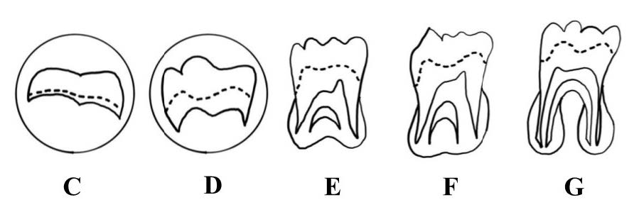
Figure 1. Drawing pictures of tooth development stages (C-G) according the Demirjian classification (Demirjian et al., 1973)
Table 2. Age estimation equations according to the Duangto et al. (Duangto et al., 2020).
|
Sex |
Equations |
|
Male |
y = 2.951 + 1.178x + 0.052x2 |
|
Female |
y = 7.097 – 0.606x + 0.214x2 |
Remarks: y = the dental age, x = the developmental score.
RESULTS
The Kappa values were 0.942 (tooth 37) and 0.916 (tooth 47) for intra-observer agreement, and 0.971 (tooth 37) and 0.858 (tooth 47) for inter-observer agreement. These values indicated almost perfect agreement according to the Landis and Koch guidelines (Landis and Koch, 1977). Moreover, the developmental stage between teeth 37 and 47 was analyzed using the Wilcoxon signed rank test. The results showed no significant differences between teeth 37 and 47 in both males (P = 0.414). and females (P = 0.052). In addition, descriptive statistics of the chronological age in stages C to G compared between males and females for teeth 37 and 47 are shown in Table 3. No significant differences in the chronological age in each developmental stage were observed, except for stage G of tooth 37, which found statistically significant differences between the males and females (P = 0.041).
Table 3. Descriptive statistics of the chronological age between males and females.
|
Tooth |
Stage |
Males (n = 112) |
Females (n = 112) |
P-value |
||
|
Mean |
SD |
Mean |
SD |
|||
|
37 |
C |
7.48 |
0.35 |
7.38 |
0.27 |
0.619 |
|
D |
8.33 |
0.79 |
8.42 |
1.04 |
0.830 |
|
|
E |
9.27 |
1.02 |
8.92 |
0.92 |
0.218 |
|
|
F |
11.22 |
0.98 |
10.78 |
1.30 |
0.203 |
|
|
G |
13.31 |
1.00 |
12.80 |
1.43 |
0.041* |
|
|
47 |
C |
7.50 |
0.32 |
7.27 |
0.22 |
0.243 |
|
D |
8.43 |
0.75 |
8.32 |
0.99 |
0.750 |
|
|
E |
9.39 |
1.11 |
9.07 |
0.91 |
0.285 |
|
|
F |
11.10 |
1.05 |
10.94 |
1.21 |
0.643 |
|
|
G |
13.28 |
1.00 |
12.93 |
1.40 |
0.153 |
|
Remarks: n = number of samples, Mean = mean age, SD = standard deviation, * statistically significant difference using the independent t test.
Finally, our results showed that the mean difference values were 0.50–0.53 years in males and 0.27–0.40 years in females. Moreover, the 95% CI showed the lower bounds and upper bounds in teeth 37 and 47 (Table 4).
Table 4. Mean difference (DA-CA) values and 95% confidence interval (95% CI) using the difference values between dental age (DA) and chronological age (CA) in teeth 37 and 47 for males and females.
|
Sex |
Tooth |
Mean DA (year) |
Mean CA (year) |
Mean DA-CA (year) |
SD |
95% CI (year) |
|
|
Lower |
Upper |
||||||
|
Male |
37 |
11.55 |
11.02 |
0.53 |
0.71 |
0.39 0.66 |
|
|
47 |
11.52 |
11.02 |
0.50 |
1.03 |
0.31 0.69 |
||
|
Female |
37 |
11.43 |
11.03 |
0.40 |
1.27 |
0.16 0.64 |
|
|
47 |
11.32 |
11.03 |
0.27 |
1.21 |
0.04 0.50 |
||
DISCUSSION
Age estimation from dental development is a popular and reliable method that is currently used in forensic sciences, clinical medicine, and clinical dentistry (Theerasopon et al., 2023). A tooth or several teeth have been used in equations for age estimation. The third molar is commonly used for age estimation (Theerasopon et al., 2022). However, this tooth has a high incidence of congenital absence (Özükoç, 2022) and may be extracted due to pain in children or adults (Al-Assadi, 2018), which makes it unsuitable for age estimation. The same applies to the first molars that are the first permanent teeth that erupt into the mouth and have the highest incidence of extraction due to dental caries (Skeie et al., 2006). On the other hand, the second molar, which is the adjacent tooth with a lower likelihood of extraction, has a development period in childhood that can be used for age estimation.
Age estimation from teeth can be done through various methods that include visual, biochemical, and radiographic methods. Age estimation through radiographic methods is a non-invasive technique. There are several classifications of radiographic methods, such as the Schour and Massler, Nolla, Moorees Fanning and Hunt, and the Demirjian method (Demirjian et al., 1973; Harris and Buck, 2002; Nandlal et al., 2014).
Age estimation based on dental development using the Demirjian method, which uses panoramic radiographs, is a precise and widely used technique that identifies developmental stages in each phase. Moreover, this method has shown excellent reproducibility for intra-observer and inter-observer agreements that were similar to this current study. Age estimation using the Demirjian method is categorized into eight stages: A to H (Demirjian et al., 1973). However, this current study did not employ stages A, B, and H since our sample population was 7–14 years old, which was the same age group used to develop the original equations by Duangto et al. The age range of 7–14 years is a period when crown formation begins but root formation is not complete. Additionally, it serves as a control variable to test the accuracy of age estimation in the Duangto et al. equations (Duangto et al., 2020).
The present study discovered that the developmental stages of teeth 37 and 47, as classified by the Demirjian method, were not significantly different between males and females. These results were consistent with the results reported in the populations of northern Thailand (Duangto et al., 2020), northern China (Guo et al., 2018), Brazil (Almeida et al., 2013), and Korea (Lee et al., 2010). Therefore, using either 37 or 47 could be used for age estimation.
The main findings of this study were the differences between dental age and chronological age for the permanent mandibular second molars as measured by the mean error in both males and females. The mean errors for tooth 37 and tooth 47 in males and females were 0.53 and 0.40 years and 0.50 and 0.27 years, respectively. Therefore, the dental ages were slightly higher than the chronological ages but were within the age range of ±1 years (Nayak et al., 2014). These results indicated the dental ages did not significantly differ from the chronological ages, which was also similar to the results found in northern Thailand (Duangto et al., 2020), Malaysia (Dewi Ardini et al., 2013), and Portugal (Fins et al., 2017).
The chronological age compared between males and females at each dental developmental stage revealed that dental development progressed faster in females than in males, which was consistent with Spanish (Feijoo et al., 2012), Brazilian (Almeida et al., 2013), northern Chinese (Guo et al., 2018), and southern African populations (Esan and Schepartz, 2019). However, it is important to note that no statistically significant differences were observed except in stage G in tooth 37.
CONCLUSION
The age estimation equations developed by Duangto et al. are valid to estimate the chronological age of both males and females in the population of southern Thailand.
ACKNOWLEDGEMENTS
The authors greatly appreciate the Dental Hospital, Faculty of Dentistry, Prince of Songkla University, Songkhla, Thailand and School of Dentistry, University of Phayao, Phayao, Thailand for facilitating support of this study.
AUTHOR CONTRIBUTIONS
Conceptualization: Pornpat Theerasopon and Phuwadon Duangto. Data acquisition: Chairat Charoemratrote, Wirin Woratanarat, Nichawan Saiwong and Namthip Netprakon. Data analysis or interpretation: Pornpat Theerasopon, Phuwadon Duangto, Wirin Woratanarat, Nichawan Saiwong and Namthip Netprakon. Drafting of the manuscript: Wirin Woratanarat, Nichawan Saiwong, Namthip Netprakon, Pornpat Theerasopon and Phuwadon Duangto. Critical revision of the manuscript: Pornpat Theerasopon and Phuwadon Duangto. Approval of the final version of the manuscript: all authors.
CONFLICT OF INTEREST
No potential conflict of interest relevant to this article was reported.
REFERENCES
Al-Assadi, A.H. 2018. Patterns and causes of teeth extraction among children attending Baghdad dental teaching hospital. Journal of International Medical Research and Health Sciences. 7(5): 88-95.
Almeida, M.S., Pontual Ados, A., Beltrão, R.T., Beltrão, R.V., and Pontual, M.L. 2013. The chronology of second molar development in Brazilians and its application to forensic age estimation. Imaging Science in Dentistry. 43(1): 1-6.
Almonaitiene, R., Balciuniene, I., and Tutkuviene, J. 2010. Factors influencing permanent teeth eruption. Part one-General factors. Stomatologija. 12(3): 67-72.
Demirjian, A., Goldstein, H., and Tanner, J.M. 1973. A new system of dental age assessment. Human Biology. 45(2): 211-227.
Dewi Ardini, Y., Sukmasari, S., and Joo Ming, C. 2013. Chronological age estimation using mandibular left permanent second molar in Malaysian children. In: 2nd MAPD Biennial Scientific Conference and Trade Exhibition, 9-11 Mar 2013. Premiera Hotel, Kuala Lumpur.
Duangto, P., Janhom, A., and Iamaroon, A. 2020. Age estimation using permanent mandibular second molar teeth in a Thai population. Australian Journal of Forensic Sciences. 53(5): 557-565.
Duangto, P., Janhom, A., Prasitwattanaseree, S., Mahakkanukrauh, P., and Iamaroon, A. 2016. Age estimation methods in forensic odontology. Journal of Dentistry Indonesia. 23(3): 74-80.
Esan, T.A. and Schepartz, L.A. 2019. The timing of permanent tooth development in a Black Southern African population using the Demirjian method. International Journal of Legal Medicine. 133(1): 257-268.
Feijóo, G., Barbería, E., De Nova, J., and Prieto, J.L. 2012. Permanent teeth development in a Spanish sample. Application to dental age estimation. Forensic Science International. 214(1-3): 213.e1-213.e6.
Fins, P., Pereira, M.L., Afonso, A., Pérez-Mongiovi, D., and Caldas, I.M. 2017. Chronology of mineralization of the permanent mandibular second molar teeth and forensic age estimation. Forensic Science, Medicine and Pathology. 13(3): 272-277.
Garn, S.M., Lewis, A.B., and Blizzard, R.M. 1965a. Endocrine factors in dental development. Journal of Dental Research. 44(1): 243-258.
Garn, S.M., Lewis, A.B., and Kerewsky, R.S. 1965b. Genetic, nutritional, and maturational correlates of dental development. Journal of Dental Research. 44(1): 228-242.
Guo, Y.C., Chu, G., Olze, A., Schmidt, S., Schulz, R., Ottow, C., Pfeiffer, H., Chen, T., and Schmeling, A. 2018. Age estimation of Chinese children based on second molar maturity. International Journal of Legal Medicine. 132(3): 807-813.
Harris, E. and Buck, A. 2002. Tooth mineralization: A technical note on the Moorrees-Fanning-Hunt standards. Dental Anthropology. 16(1): 15-20.
Hilgers, K.K., Akridge, M., Scheetz, J.P., and Kinane, D.E. 2006. Childhood obesity and dental development. Pediatric Dentistry Journal. 28(1): 18-22.
Landis, J.R. and Koch, G.G. 1977. The Measurement of observer agreement for categorical data. Biometrics. 33(1): 159-174.
Lee, S.S., Byun, Y.S., Park, M.J., Choi, J.H., Yoon, C.L., and Shin, K.J. 2010. The chronology of second and third molar development in Koreans and its application to forensic age estimation. International Journal of Legal Medicine. 124(6): 659-665.
Liu, H., Deng, H., Cao, C.F., and Ono, H. 1998. Genetic analysis of dental traits in 82 pairs of female-female twins. Chinese Journal of Dental Research. 1(3): 12-6.
Nandlal, B., Patil, D. and Shanthraj, R. 2014. Estimation of dental age by Nolla's method using Orthopantomographs among rural free residential school children. International Journal of Medical Research and Health Sciences. 3(2): 273-277.
Nayak, S.D., Geoge, R., Shenoy, A., and Shivapathasundaram, B. 2014. Age estimation in forensic dentistry-A review. International Journal of Science and Research. 3(4): 333-338.
Olze, A., Reisinger, W., Geserick, G., and Schmeling, A. 2006. Age estimation of unaccompanied minors. Part II. Dental aspects. Forensic Science International. 159: S65-S67.
Özükoç, C. 2022. Prevalence of congenital tooth deficiency: Retrospective cross-sectional study. Eastern Journal of Medicine. 27(1): 182-186.
Psoter, W., Gebrian, B., Prophete, S., Reid, B., and Katz, R. 2008. Effect of early childhood malnutrition on tooth eruption in Haitian adolescents. Community Dentistry and Oral Epidemiology. 36(2): 179-189.
Schmeling, A., Geserick, G., Reisinger, W., and Olze, A. 2007. Age estimation. Forensic Science International. 165(2-3): 178–81.
Skeie, M.S., Raadal, M., Strand, G.V., and Espelid, I. 2006. The relationship between cariesin the primary dentition at 5 years of age and permanent dentition at 10 years of age - a longitudinal study. International Journal of Paediatric Dentistry. 16(3): 152-160.
Srisuwannachit, K. 2020. Age assessment of child laborers. Thai Journal of Public Health. 50(1): 99-110.
Theerasopon, P., Charoemratrote, C., and Duangto, P. 2023. Age estimation using the Demirjian and Willems methods in a southern Thai population. Natural and Life Sciences Communications. 22(4): e2023058.
Theerasopon, P., Tiansuwan, K., Srichaitan, N., Norkaew, S., Charoemratrote, C., Srimaneekarn, N., and Duangto, P. 2022. Testing the accuracy of an age estimation method using radiographs of permanent mandibular third molar teeth in a Thai population. Chiang Mai University Journal of Natural Science. 21(3): e2022045.
OPEN access freely available online
Natural and Life Sciences Communications
Chiang Mai University, Thailand. https://cmuj.cmu.ac.th
Pornpat Theerasopon1, Wirin Woratanarat2, Nichawan Saiwong2, Namthip Netprakon2, Chairat Charoemratrote3, and Phuwadon Duangto4, *
1 Department of Orthodontics, School of Dentistry, University of Phayao, Phayao 56000, Thailand.
2 School of Dentistry, University of Phayao, Phayao 56000, Thailand.
3 Department of Preventive Dentistry, Faculty of Dentistry, Prince of Songkla University, Songkhla 90112, Thailand.
4 Department of Anatomy, School of Medical Sciences, University of Phayao, Phayao 56000, Thailand.
Corresponding author: Phuwadon Duangto, E-mail: pete_anatomy@hotmail.com
Total Article Views
Editor: Anak Iamaroon,
Chiang Mai University, Thailand
Article history:
Received: March 9, 2024;
Revised: April 22, 2024;
Accepted: April 22, 2024;
Online First: May 2, 2024